


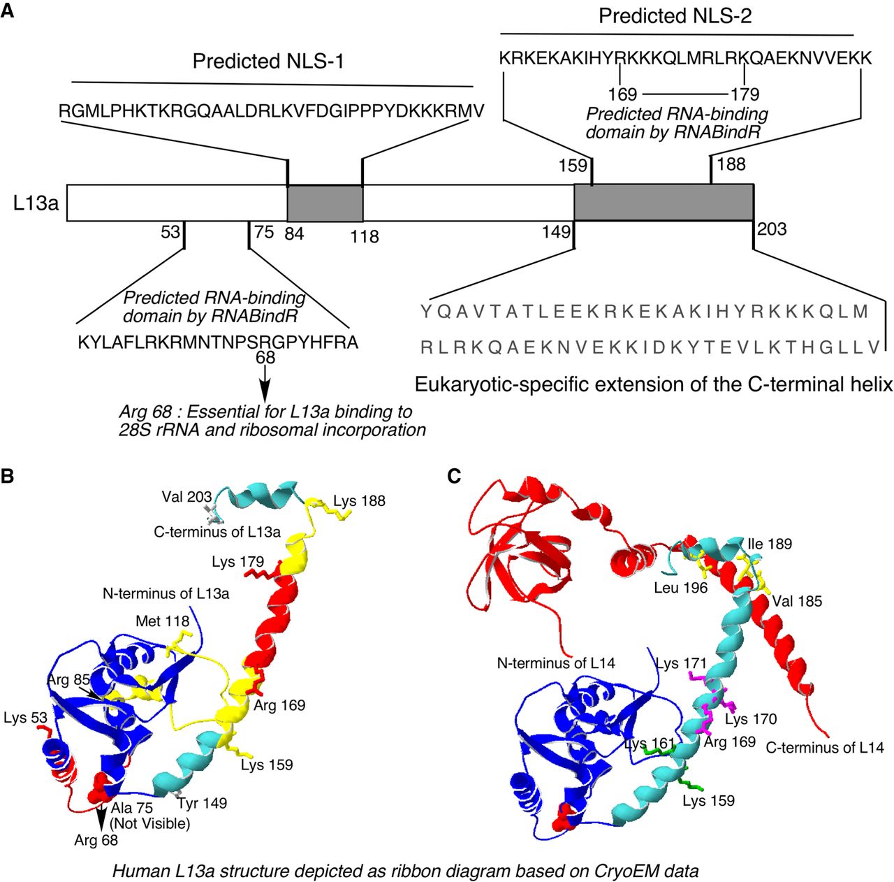
Putative NLSs, RNA binding sites, and structure of ribosomal protein L13a. (A) Schematic diagram of the human L13a amino acid sequence with predicted NLSs (Arg84 to Met118 and Lys159 to Lys188) and rRNA-binding sites (Lys53 to Ala75 and Arg169 to Lys179) indicated. Arg68, a residue experimentally verified as essential for rRNA-binding and ribosomal incorporation, is also indicated. The eukaryote-specific L13a carboxy-terminal extension spans residues Trp149 to Val203. (B) Human L13a structure depicted as a ribbon diagram based on the CryoEM structure of the human ribosome at 3.6 Å resolution (Protein Data Bank code 5T2C). The conserved globular core domain is shown in dark blue. Predicted NLSs (Arg84 to Met118 and Lys159 to Lys188) are shown in yellow. Predicted RNA-binding sites (Lys53 to Ala75 and Arg169 to Lys179) are shown in red. The eukaryote-specific carboxy-terminal extension (Tyr149 to Val203) is shown in sky blue. Side chains are shown as sticks for Lys53, Lys159, Lys179, Lys188, Ala75, Arg85, Arg169, Met 118, Tyr149, and Val203. (C) Modeling of the interaction between the carboxy-terminal helices of human L13a and L14 proteins. L13a (blue and sky blue) and L14 (red) protein structures are depicted as ribbon diagrams based on the CryoEM structure of the human ribosome at 3.6 Å resolution (Protein Data Bank code 5T2C). Side chains of the L13a residues experimentally determined to affect L13a ribosomal incorporation (Arg68, Lys159, Lys161, Arg169, Lys170, and Lys171) are shown. The van der Waals radius of Arg68 (also essential for L13a ribosomal incorporation) is shown in red. Side chains of L13a residues Val185, Ile189, and Leu196 interacting with the L14 carboxy-terminal helix are shown in yellow.




