


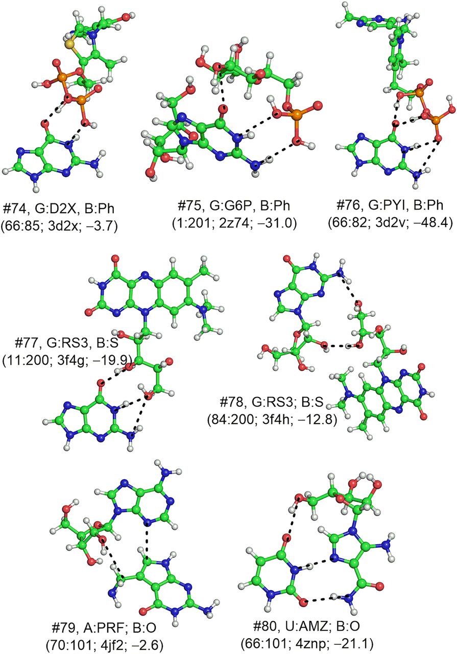
B3LYP/6-31G(d,p) optimized geometries of base:phosphate (B:Ph), base:sugar (B:S), and base:other (B:O) type of base:ligand interactions. Structure number (#), base:ligand interaction identity and interaction type (B:Ph, B:S or B:O) are provided against each structure for identification from tables. Base-pairing interaction code and PDB code of the crystal structure used for optimization, as well as the MP2/aug-cc-pVDZ interaction energy (kcal/mol) are provided for each structure in parentheses. Hydrogen bonding donor–acceptor (D–A) distance (Å), acceptor-hydrogen (A–H) distance (Å), and D–H–A (deg) is provided for each hydrogen bond (dotted lines) in Supplemental Table S10. For structures #74, #78, and #79, only the positions of hydrogen atoms were optimized, whereas full optimization was carried out for all other structures. Structures #74 and #76 contain positively charged ligand moieties.




