


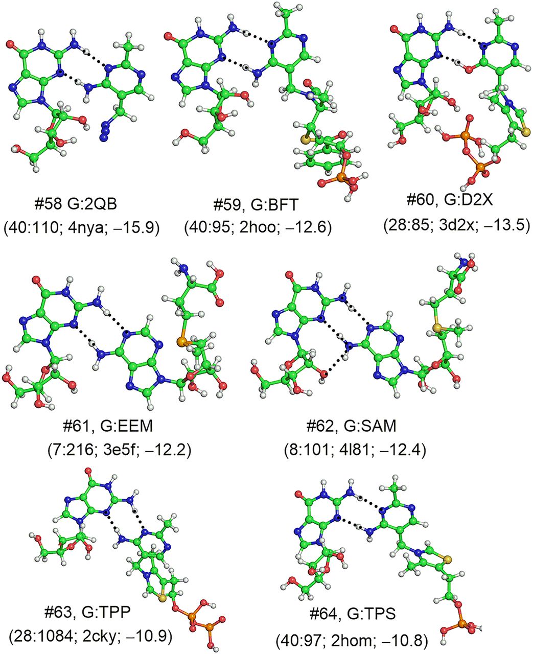
B3LYP/6-31G(d,p) optimized geometries of base:base (B:B) type of base:ligand interactions that resemble the trans Sugar–Watson Crick (cSW) RNA base pairs. Structure number (#) and base:ligand interaction identity are provided against each structure for identification from tables. Base-pairing interaction code and PDB code of the crystal structure used for optimization, as well as the MP2/aug-cc-pVDZ interaction energy (kcal/mol) are provided for each structure in parentheses. Hydrogen bonding donor–acceptor (D–A) distance (Å), acceptor–hydrogen (A–H) distance (Å), and D–H–A (deg) are provided for each hydrogen bond (dotted lines) in Supplemental Table S9. For structure #60, only the positions of hydrogen atoms were optimized, whereas full optimization was carried out for all other structures. Structures #60 to #64 contain positively charged ligand moieties.




