


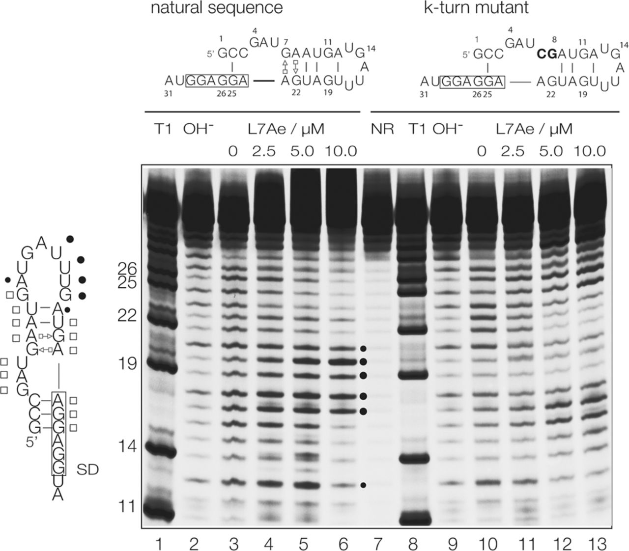
Analysis of conformational change in the A. fulgidus 5′-UTR RNA induced by the binding of AfL7Ae protein using in-line probing (Soukup and Breaker 1999). The sequences of the two RNA species studied are shown at the top, drawn in their potential stem–loop conformations. The boxed regions are the ribosome binding site. The mutant sequence (right) has two sequence changes (highlighted in bold) in the G:A base pairs that will prevent k-turn formation. Tracks 1 and 8 contain RNA cleaved by T1 nuclease that cuts 3′ to G nucleotides to provide a reference frame (sequence positions shown on the left). Tracks 2 and 9 contain natural-sequence RNA subject to hydroxide cleavage to generate cleavage through the RNA. Track 7 contains RNA not subjected to any degradation procedure, i.e., full-length RNA. Tracks 3 through 6 and 10 through 13 contain the results of in-line probing in the presence of 0, 2.5, 5, and 10 µM AfL7Ae for natural and mutant RNA, respectively. The black circles drawn on the fluorogram denote positions of reactivity in the natural 5′-UTR RNA in the presence of L7Ae. The schematic on the left shows the sequence of the natural 5′-UTR drawn in its stem–loop conformation. Filled circles denote positions of reactivity and open squares denote positions of protection, both in the presence of L7Ae.




