


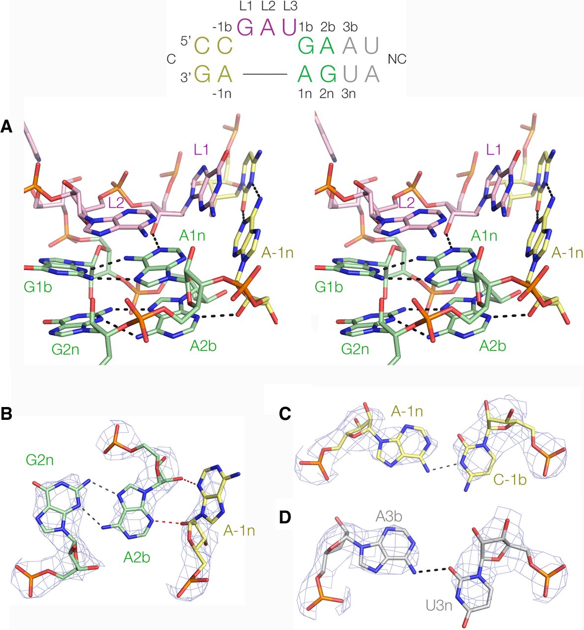
The structure of the A. fulgidus 5′-UTR k-turn in complex with L7Ae. The sequence and nucleotide nomenclature for the k-turn is shown at the top. (A) A parallel-eye stereoscopic image of the core of the k-turn. The RNA adopts the conformation of a standard, simple k-turn with two trans-sugar-Hoogsteen G:A base pairs and cross-strand hydrogen bonds from the O2′ groups of L1 and −1n ribose to the nucleobases of A1n and A2b, respectively. B through D show details from the structure with electron density from the 2Fo−Fc map contoured at 2.5 σ. (B) The A2b:G2n base pair and its interaction with A-1n. A-1n N3 accepts a hydrogen bond from A2b O2′, and its O2′ donates one to A2b N3. The A2b:G2n base pair is associated by two hydrogen bonds. This k-turn is therefore a standard N3 structure (Daldrop and Lilley 2013). (C) The C-1b:A-1n base pair. This forms a cis base pair associated by a single hydrogen bond from A-1n N6 to C-1b N3. (D) The A3b:U3n base pair. This forms a trans base pair associated by a single hydrogen bond from A3b N6 to U3n O2.




