


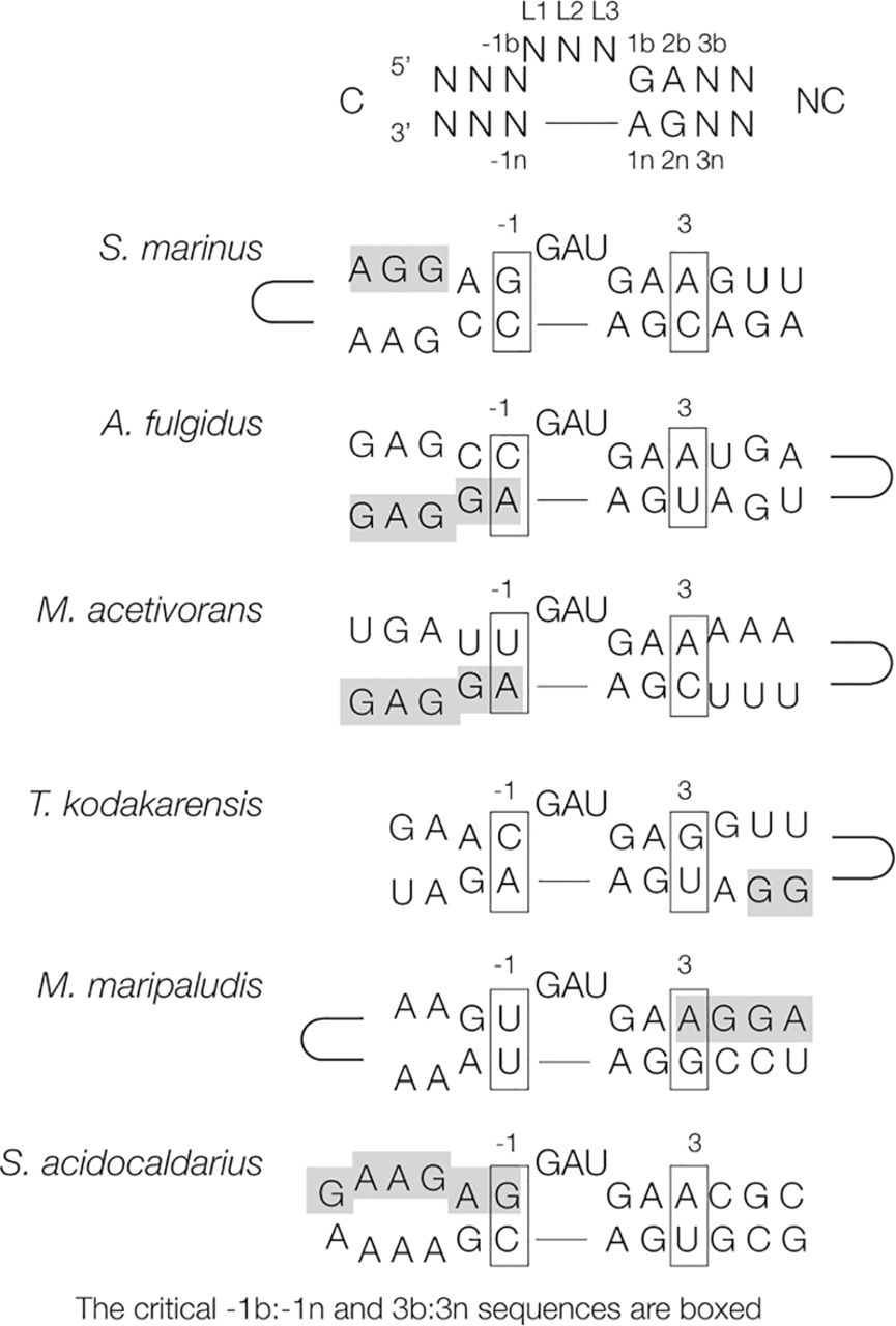
FIGURE 1.
Potential k-turn sequences occurring in the 5′-UTR sequences of the mRNA of archaeal l7ae genes. Each sequence is drawn in the form of a hairpin loop containing a putative k-turn structure, with the critical −1b:−1n and 3b:3n sequences boxed. The dependence of ion-dependent folding on the 3b:3n sequence is summarized in Supplemental Figure S1. The Shine–Dalgarno (ribosome binding site) sequences are shaded. The locations of the terminal loops are indicated schematically. The nomenclature of sequence positions for a standard k-turn is shown at the top. Sequence information was taken from Daume et al. (2017).




