


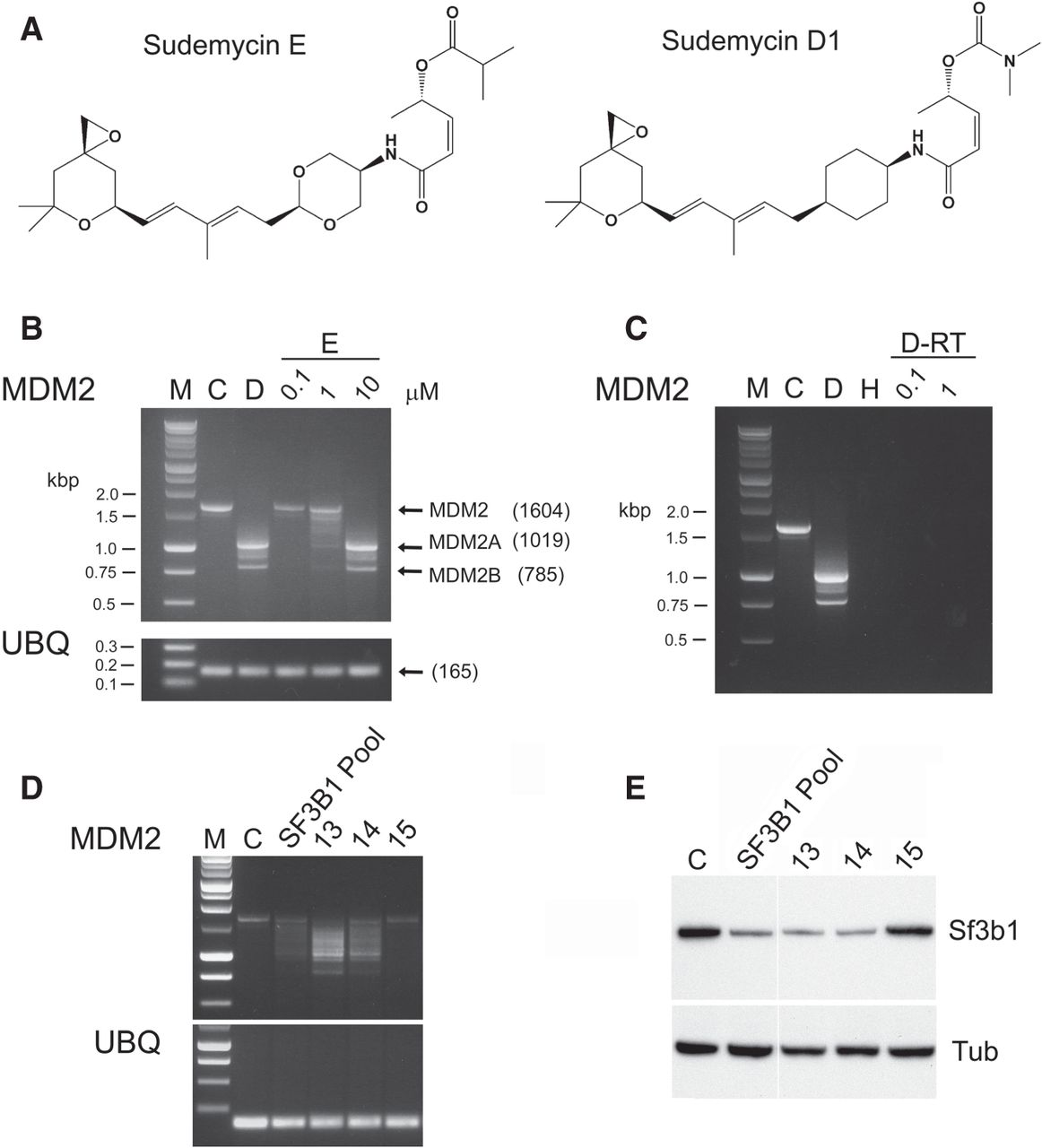
Aberrant splicing induced by sudemycin D1. (A) The chemical structure of sudemycin D1 and E. (B) Sudemycin D1 and E result in AS of MDM2 following exposure of Rh18 cells. M—Molecular weight markers; C—untreated cells; D—cells treated with 1 µM sudemycin D1 for 8 h; 0.1, 1, 10—cells treated with the corresponding concentration (µM) of sudemycin E for 8 h. Ubiqutin (UBQ) was used as a control for these analyses. Size markers are indicated in kbp with individual transcript sizes in bp (in parentheses). (C) RT/PCR controls to demonstrate that no DNA (H), and samples lacking reverse transcriptase (RNA obtained from cells treated with either 0.1 µM [D-RT 0.1] or 1 µM [D-RT 1] sudemycin D1), do not yield signals following PCR. (D) PCR analysis of MDM2 transcripts following siRNA treatment of Rh18 cells. M—Molecular weight markers; C—untreated cells. SF3B1 Pool—pool of four siRNAs targeting SF3B1; 13, 14 and 15—individual siRNAs present within the SF3B1 Pool. Ubiqutin (UBQ) was used as a control for these analyses. (E) Western analyses demonstrating the loss of SF3B1 protein in Rh18 cells following treatment with pooled or individual siRNAs that target the RNA encoding this protein. Tubulin (Tub) was used as a loading control.




