


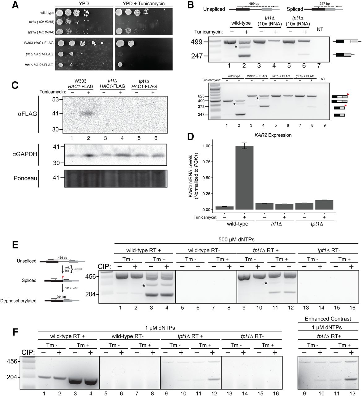
UPR-related phenotypes of RNA repair mutants. (A) Growth assay of RNA repair mutants on UPR-inducing media. Yeast cells (wild-type, trl1Δ, and tpt1Δ) were serially diluted and spotted onto rich media (YPD) and tunicamycin-containing media (80 ng/mL tunicamycin) to induce the UPR. Plates were imaged after 3 d of growth at 30°C. Wild-type and tpt1Δ cell growth is unaffected by tunicamycin, whereas trl1Δ cells fail to grow on media containing tunicamycin. Serial dilution growth assays for cells of the same genetic background as above, but with C-terminal FLAG tags on Hac1, are shown below. (B) Analysis of HAC1 splicing in RNA repair mutants. Total RNA from untreated and tunicamycin-treated wild-type, trl1Δ, and tpt1Δ cells was analyzed by RT-PCR using primers specific for HAC1, producing products at 499 bp (unspliced HAC1) and 247 bp (spliced HAC1). A no-template (NT) control is shown in lane 7. The proportion of spliced HAC1 upon tunicamycin treatment increases in wild-type (lanes 1, and 2) and tpt1Δ cells (lanes 5 and 6), and spliced HAC1 is visible in RNA from both cells (lanes 2 and 6, 247 bp). Spliced HAC1 is undetectable in trl1Δ cells (lane 4) upon tunicamycin treatment (lanes 3 and 4), owing to the inability of trl1Δ cells to ligate HAC1 exons. Asterisk marks an unknown PCR product dependent on tunicamycin treatment. Cells with C-terminal FLAG tags of Hac1 were also analyzed for splicing in the same manner (below) with a no-template (NT) control in lane 9. Reactions lacking reverse transcriptase (RT−) were negative for amplification (data not shown). (C) Hac1 protein levels in RNA repair mutants. Whole cell lysates were prepared from wild-type, trl1Δ, and tpt1Δ cells expressing C-terminal Hac1-FLAG and grown in the presence and absence of tunicamycin. Lysates were analyzed by SDS–PAGE and nitrocellulose transfer followed by Ponceau S staining and cross-reaction with anti-FLAG and anti-GAPDH antibodies. Scale to the left is nominal molecular mass of a protein ladder (kDa); the expected mass of Hac1-FLAG is 31 kDa. Hac1-FLAG is detected in wild-type cells upon tunicamycin addition, but is undetectable in trl1Δ and tpt1Δ cells. (D) Induction of the UPR-responsive KAR2 gene in RNA repair mutants. Amounts of KAR2 mRNA (normalized to PGK1 mRNA abundance) were measured by RT-qPCR in wild-type, trl1Δ, and tpt1Δ cells in the presence and absence of tunicamycin. Error bars are 95% confidence intervals, n = 3. Relative abundance of KAR2 mRNA increased 20-fold in wild-type cells treated with tunicamycin, whereas the corresponding levels of KAR2 did not increase in trl1Δ and increased 1.4-fold in tpt1Δ cells. (E) Detection of ligated and 2′-phosphorylated HAC1 mRNA. Total RNA was treated with calf intestinal phosphatase (CIP) to remove 2′-phosphates (diagram) and reverse-transcribed using HAC1-specific primer under high (500 µM) concentrations of dNTPs. The cDNA products were PCR amplified, yielding products for unspliced (456 bp) and spliced (204 bp) HAC1 mRNA. Using high dNTP concentrations, we find that splicing of HAC1 in wild-type cells increases upon tunicamycin treatment (compare lanes 1 and 2 versus 3 and 4), similar to B, but is unaffected by CIP treatment (compare lanes 1 versus 2, and 3 versus 4). Likewise, we find that spliced HAC1 mRNA in tpt1Δ cells increases in response to tunicamycin (cf. lanes 9 and 10 versus lanes 11 and 12), albeit to a lesser extent than wild-type cells, and is unaffected by CIP treatment (cf. lanes 9 versus 10 and lanes 11 versus 12). An asterisk marks an unknown PCR product dependent on tunicamycin treatment. (F) Detection of ligated and 2′-phosphorylated HAC1 mRNA. Using low (1 µM) dNTP concentrations, we find that spliced HAC1 is preferentially amplified in wild-type cells over unspliced HAC1 (lanes 1–4). The abundance of spliced HAC1 mRNA from wild-type cells increases in response to tunicamycin but is unaffected by CIP treatment (cf. lanes 1 versus 2 and 3 versus 4). RT-PCR analysis of HAC1 mRNA from tpt1Δ cells reverse transcribed under low dNTP concentrations shows both unspliced and spliced forms of HAC1 mRNA, and spliced HAC1 mRNA increases in response to tunicamycin treatment (cf. lanes 10 and 12). However, in contrast to wild-type, amplification of spliced HAC1 mRNA from tpt1Δ is strongly dependent on prior treatment with CIP. In the absence of tunicamycin and CIP treatment, spliced HAC1 mRNA is undetectable, whereas treatment with CIP enables reverse transcription (cf. lane 9 to lane 11; see panel with enhanced contrast to the right). Similarly, the abundance of spliced HAC1 mRNA from tpt1Δ cells increases in response to tunicamycin, and its abundance is further increased upon CIP treatment (lane 11 versus 12).




