


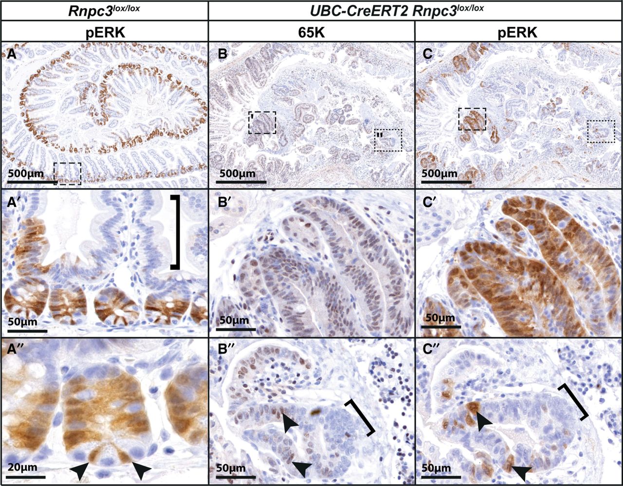
pERK expression is concordant with 65K expression in the proliferative cells of intestinal crypts and in recovering regions of the epithelium. (A) Immunohistochemistry of pERK1/2 (pERK) reveals staining in the BrdU-positive compartment (cf. Fig. 5C,D) of crypts (boxed region in A is shown at higher magnification in A′ and A″). Meanwhile, pERK staining is absent from the terminally differentiated cells of the villi (bracket A′). The crypt base columnar (CBC) cells (arrowheads A″) strongly express pERK and can be clearly seen wedged between the large, granular, terminally differentiated Paneth cells, which are pERK negative. (B) Six days following tamoxifen treatment to induce recombination of Rnpc3lox alleles, the structural organization of the small intestinal epithelium is lost, save for discontinuous patches of cells that still express 65K due to incomplete recombination of Rnpc3lox alleles (B′ and arrows B″). (C) These same cells also show robust nuclear and cytoplasmic pERK expression (C is a serial section of B). The left box in B is magnified in panels B′ and C′. Regions predominantly lacking 65K (right box in B) also fail to express pERK (see brackets in magnified panels B″ and C″). Arrows denote individual cells in B and C that are both 65K- and pERK-positive (note that the 65K staining is nuclear only, whereas the pERK staining is found in the nucleus and cytoplasm). Scale bars in A, B, and C = 500 µm. Scale bars in A′, B′, C′, B″, and C″ = 50 µm. Scale bar in A″ = 20 µm.




