


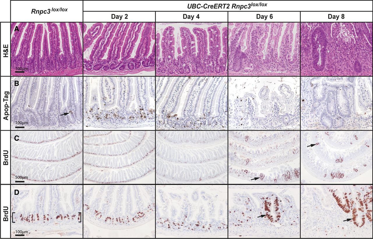
Loss of 65K induces apoptosis of epithelial cells in small intestinal crypts. The impact of acute global 65K deficiency on the integrity of the small intestine was studied in tamoxifen-treated Rnpc3lox/lox adult mice (8–12 wk) harboring an inducible UBC-CreERT2 allele. (A) Imaging of hematoxylin and eosin (H&E)-stained sections of small intestine from UBC-CreERT2;Rnpc3lox/lox mice revealed progressive epithelial damage from 4 d post-tamoxifen treatment, characterized by loss of crypt structure, shortening of villi, and invasion of inflammatory cells. Enlarged, regenerative crypts 6 d and 8 d post-tamoxifen treatment are indicative of a wound healing response. (B) Apo-tag (brown staining) identifies cells undergoing apoptosis. Such cells are rare in the unrecombined epithelium (arrow), but increase markedly in number in UBC-CreERT2;Rnpc3lox/lox mice 2 d and 4 d post-tamoxifen treatment. The apoptotic cells are found mainly in the crypts. (C) At 2, 4, 6, and 8 d post-tamoxifen treatment, mice were injected with BrdU to mark cells in the S-phase of the cell cycle, prior to being euthanized 2 h later. Low magnification images of BrdU-positive cells (brown nuclei) throughout the small intestine highlight the location of proliferative cells in the crypts; such cells are absent from the villi. Scale bar = 500 µm. Upon recombination, BrdU-positive cells are fewer in number and more dispersed, particularly 4 d after tamoxifen treatment. Areas of regenerating proliferating cells are observed from 6- to 8-d post-tamoxifen treatment (arrows). (D) Higher magnification views highlighting proliferative cells in control crypts (brackets) and at sites of regeneration in Rnpc3-depleted intestinal epithelium (arrows). Scale bars in A, B, D = 100 µm (applies to all images in the row).




