


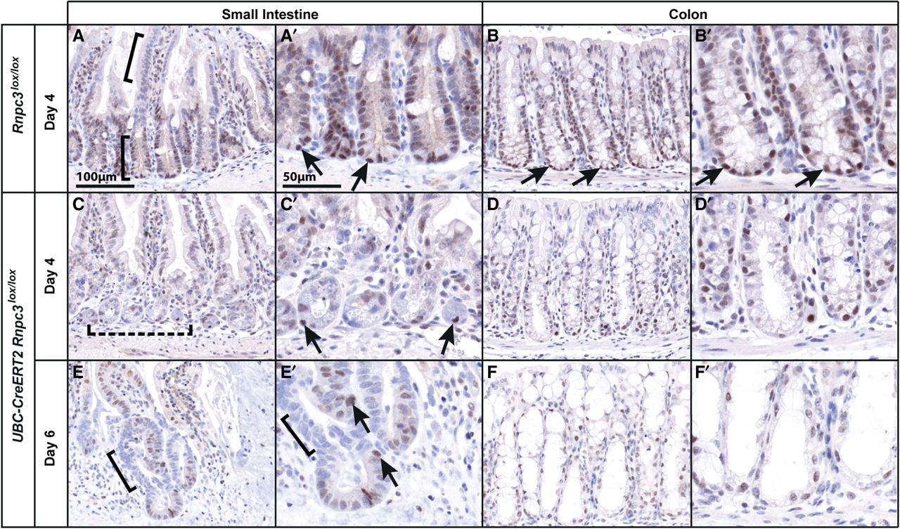
65K is expressed in the nuclei of cells in intestinal crypts and is lost following induced Rnpc3 deletion. (A,A′) Immunostaining with 65K antibody highlights positive nuclei at the base of the crypts of the small intestine (A, lower bracket) and colon (B,B′) with weak to absent expression in cells of the small intestinal villi (A, upper bracket) and in the lamina propria. Black arrows denote cells with 65K-positive nuclei in the stem cell compartment of the crypts. (C,D) Four days after tamoxifen (TMX) treatment of UBC-CreERT2;Rnpc3lox/lox mice, 65K protein expression is decreased in the small intestine (C, dashed bracket; magnified in C′) and colon (D,D′). The small intestine epithelial structure is disorganized with shorter villi. (E,F) Six days after TMX treatment, some areas of the small intestinal (E, bracket; magnified in E′) and colonic epithelium (F,F′) in UBC-CreERT2;Rnpc3lox/lox mice have completely lost 65K expression, coinciding with widespread epithelial disruption. Meanwhile, foci of regenerating crypt-like structures arise from cells in which complete recombination has not occurred and 65K protein is still evident (E′, arrows). A′–F′ show regions of interest at higher magnification. Scale bar in A, B, C, D, E, and F = 100 µm. Scale bar in A′, B′, C′, D′, E′, and F′ = 50 µm.




