


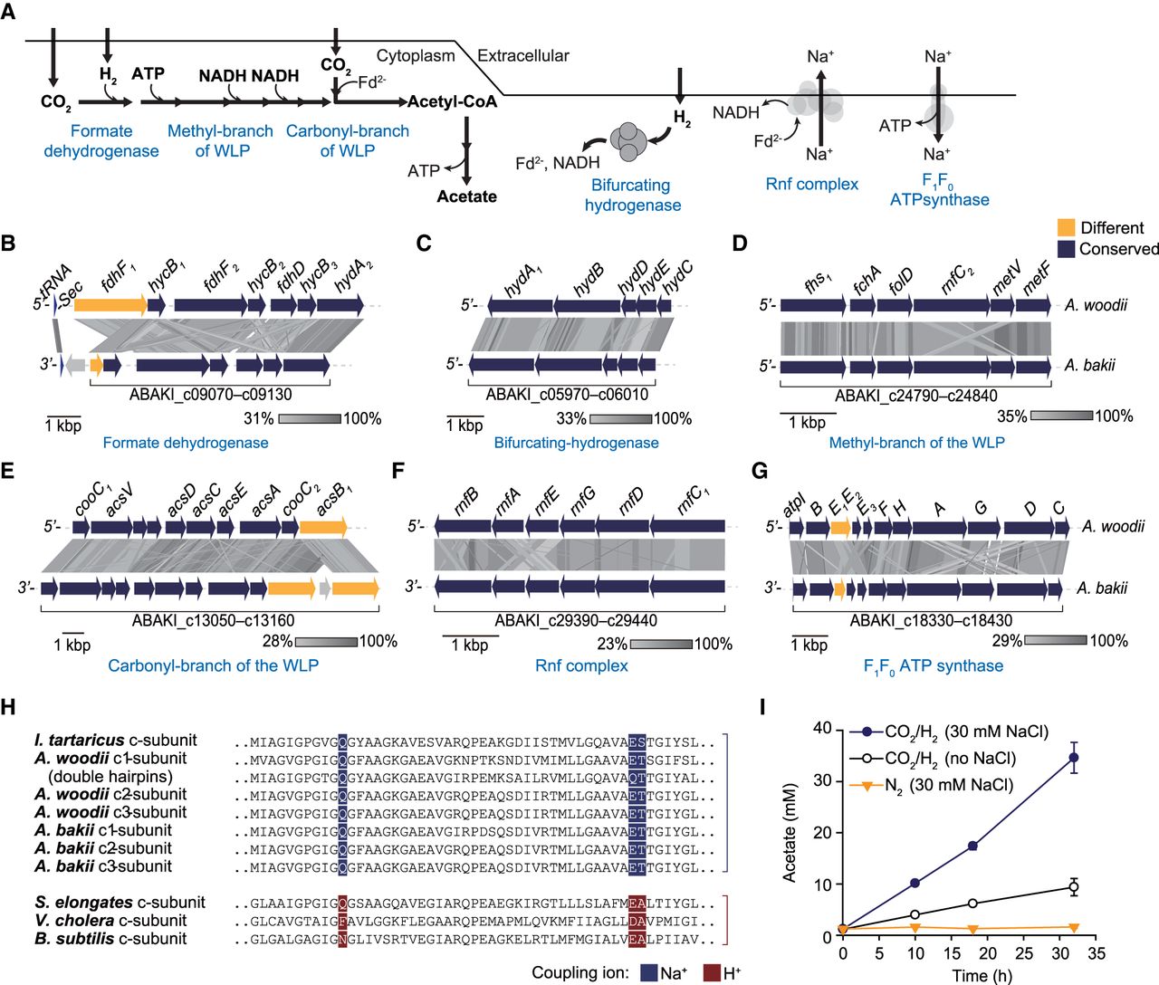
The WLP of A. bakii. (A) Schematic representation of the putative acetogenesis in A. bakii. (B–G) Synteny analysis of major gene clusters involved in acetogenesis between A. bakii and A. woodii. Comparisons were performed using tBLASTx within Easyfig (Sullivan et al. 2011), and each arrow indicates a gene. Genetic conservation is represented by a color. (H) Comparison of the sequences of the c-subunits of ATP synthase. These c-subunits assembled into homo- or heteromeric c-rings and play a direct role in ion translocation across the membrane. To determine the Na+/H+ binding motif, the c-subunit sequences of F1F0-type ATP synthase complex were aligned. The Na+/H+ binding motif, either predicted (A. bakii) or known (other strains), is highlighted in color in each case. Used c-subunit sequences were obtained by the following accession numbers or locus_tags: Ilyobacter tartaricus (Q8KRV3), Acetobacterium woodii (Awo_c02160 – c02180), Acetobacterium bakii (ABAKI_c18390–c18410), Synechococcus elongates (YP_399351), Vibrio cholera (Q9KNH0), and Bacillus subtilis (P37815). (I) Resting cell assay was performed with or without 30 mM NaCl. The whole cell of A. bakii (0.25 mg/mL) was incubated with 50 mM imidazole, 20 mM MgSO4, 20 mM KCl, 4.4 µM resazurin, 4 mM DTE at pH 7.0. The gaseous atmosphere was H2–CO2 (80:20, v/v, 200 kPa) or N2 (200 kPa). All values are means obtained from two independent experiments.




