


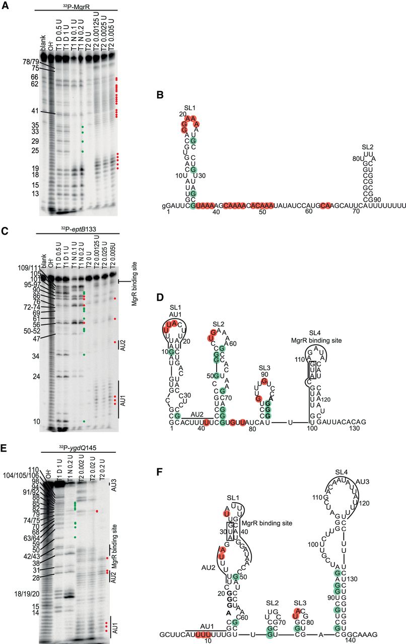
Secondary structures of E. coli MgrR sRNA and 5′-terminal fragments of mRNAs eptB and ygdQ. (A) The probing of 32P-MgrR structure with RNases indicated above the lanes. (B) The secondary structure of MgrR predicted by RNAstructure software based on data from probing experiments in A and in Supplemental Figure S1A. (C) The probing of 32P-eptB133 mRNA structure with RNases indicated above the lanes. (D) The secondary structure of eptB133 mRNA predicted by RNAstructure software based on data from probing experiments in C and in Supplemental Figure S1B. (E) The probing of 32P-ygdQ145 mRNA structure with RNases indicated above the lanes. (F) The secondary structure of ygdQ145 mRNA predicted by RNAstructure software based on data from probing experiments in E and in Supplemental Figure S1C. Symbols T1 D and T1 N denote probing with RNase T1 in denaturing or native conditions, respectively, while T2 denotes probing with RNase T2. The numbers to the left indicate positions of guanosine residues. Blank denotes untreated control and OH− denotes formamide ladder. Positions of the residues susceptible to degradation by RNase T2, Nuclease S1 (Supplemental Fig. S1A), or RNase T1 in native conditions were constrained as single-stranded (red circles) in secondary structure prediction using RNAstructure software (Reuter and Mathews 2010), while positions protected from RNase T1 cleavage in native conditions as compared to the denaturing conditions were constrained as double-stranded (green circles). MgrR binding sites and the locations of AU-rich sequence motifs are marked with lines in D and F. The AUG start codon is boxed and SD sequences are marked in bold font.




