


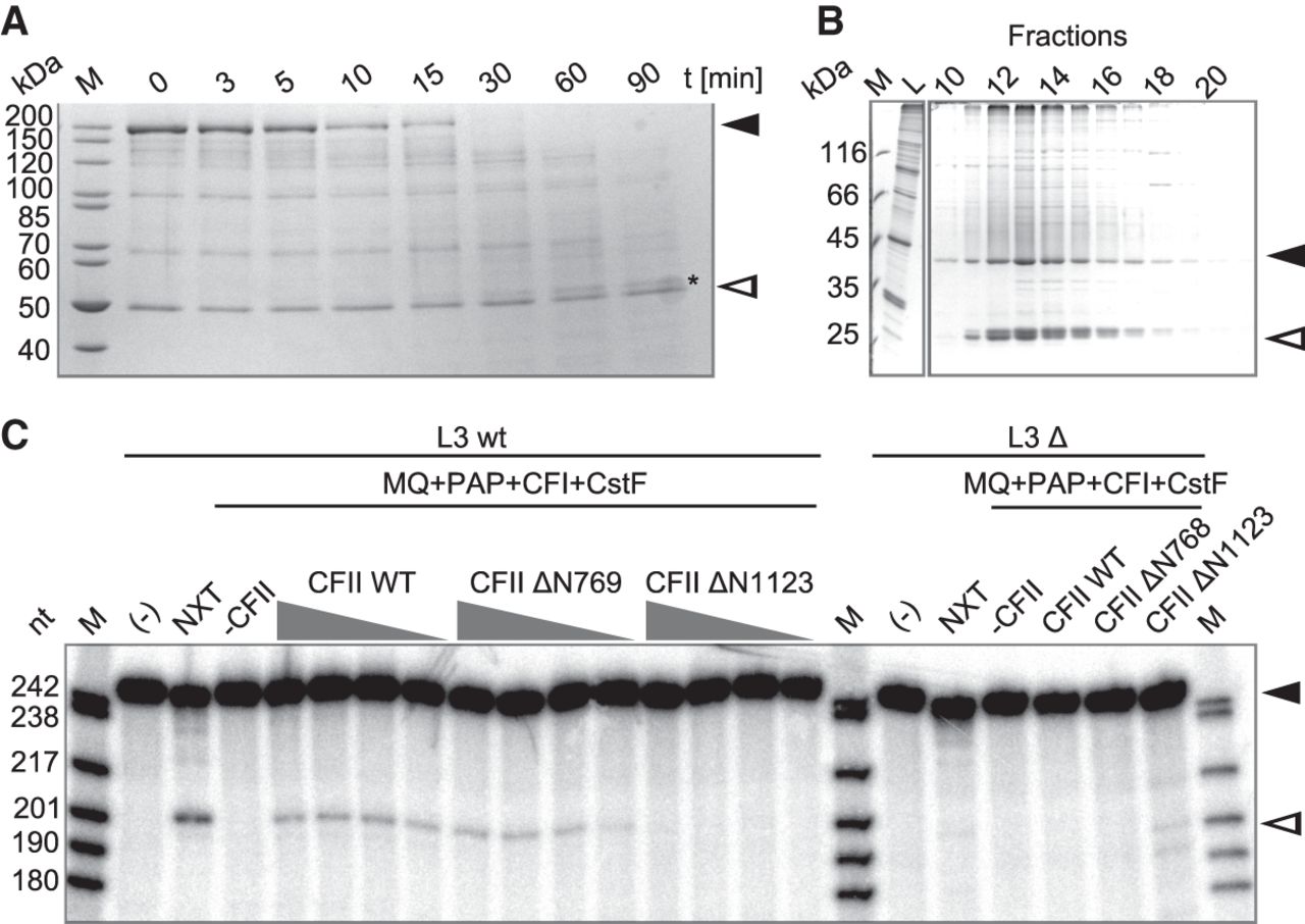
Domains of hPcf11. (A) Trypsin digestion identifies a stable C-terminal hPcf11 fragment. Human Pcf11 was digested with trypsin for the times indicated (see Materials and Methods). Proteins were analyzed on an SDS-polyacrylamide gel and stained with Coomassie. Black and white arrowheads indicate full-length hPcf11 and hClp1, respectively, and the asterisk marks the stable hPcf11 fragment. (B) The C-terminal region of hPcf11 containing the hClp1 interaction sequence flanked by the zinc fingers suffices for stable hClp1 binding. His-tagged hClp1 and untagged hPcf11ΔN1339 were coexpressed and purified via Ni-NTA and MonoQ chromatography. The figure shows an analysis of the MonoQ peak fractions by SDS-PAGE and Coomassie staining. Black and white arrowheads indicate hClp1 and the hPcf11 fragment, respectively. Identity and completeness of the hPcf11 fragment were confirmed by LC/MS/MS. (C) A C-terminal hPcf11 fragment starting with the FEGP repeats supports pre-mRNA cleavage. Cleavage assays were performed with the L3 RNA and mutant control and proteins as described in the legend to Figure 2B. Controls are as in Figure 2B. CF II ΔN769 and ΔN1123 refers to the heterodimers reconstituted with the respective hPcf11 mutants. Black and white arrowheads indicate substrate RNA and 5′ cleavage product, respectively.




