


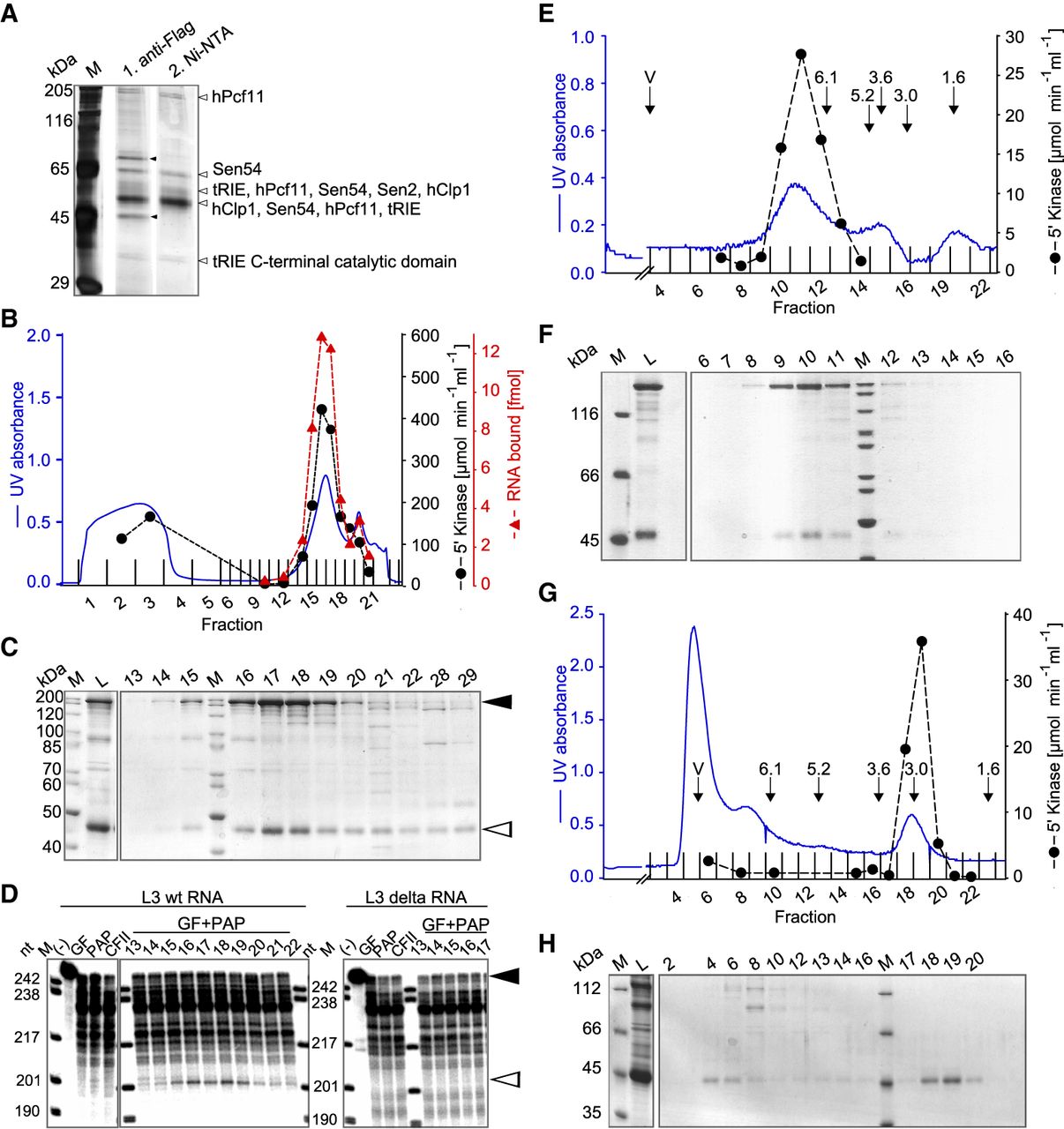
CF II is a heterodimer of hClp1 and hPcf11 and copurifies with RNA 5′ kinase, RNA binding, and 3′ cleavage activity. (A) His-FLAG-tagged hClp1 was affinity purified from a stable cell line by two consecutive purification steps on FLAG beads and Ni2+ beads. The silver-stained SDS gel shows the eluates from the first (middle lane) and second purification step (right lane). The two bands marked in the FLAG eluate are PRMT5 and MEP50, known FLAG-binding proteins. Sen2, Sen54, and tRIE are subunits of the tRNA splicing endonuclease. (B) MonoQ column profile of his-tagged hClp1 and hPcf11 coexpressed in baculovirus-infected insect cells (see Material and Methods). RNA binding was measured by nitrocellulose filter binding assays with 0.5 µL of a 1:100 dilution of the fractions indicated and 100 fmol of SV40 late RNA (see Materials and Methods). Kinase activity was assayed as described in Materials and Methods. (C) Fractions of the column shown in B were analyzed by SDS-PAGE. Arrowheads indicate hPcf11 and hClp1. (L) Load of column. (D) Fractions of the column shown in B were assayed for reconstitution of 3′ cleavage as described in Materials and Methods. Assays contained poly(A) polymerase (PAP) and a fraction (GF) generated by ammonium sulfate precipitation of HeLa cell nuclear extract and Superose 6 gel filtration; this fraction contained all factors required for cleavage except PAP and CF II. Reactions were complemented with 2 µL of the MonoQ column fractions and were carried out with wild-type L3 RNA or L3Δ RNA with a point mutation in the AAUAAA signal. With both RNAs, the first four lanes following the marker lane show negative controls, as indicated. Black and white arrowheads indicate RNA substrates and specific cleavage products. AAUAAA- and CF II-independent partial degradation of most of the substrate is due to nuclease activity in GF (compare control lanes). (E) Gel filtration of CF II on a Superose 6 column. Arrows indicate the void volume (V) and the elution peaks of marker proteins with their Stoke's radii (see Materials and Methods). (F) SDS-PAGE of the column fractions of E. (G) Superdex 200 gel filtration column profile of His-tagged hClp1 expressed in baculovirus-infected cells (see Materials and Methods). RNA kinase activity was measured as described in Materials and Methods. The void volume (V) and peak positions of marker proteins with their Stoke's radii are indicated. (H) SDS-PAGE of the column fractions of G.




