


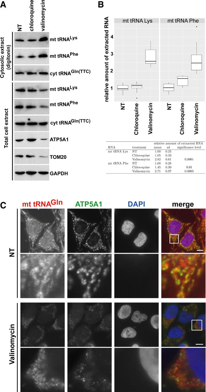
Induction of autophagy increases soluble cytosolic mt tRNA levels. (A) Accumulation of mt tRNAs in the cytosol of HeLa cells undergoing autophagy. Cytosolic and total cellular extracts were prepared from HeLa cells nontreated (NT) or treated with chloroquine and valinomycin by mild digitonin extraction or sonication, respectively. The levels of mt tRNAs Lys and Phe and the mitochondrial proteins ATP5A1 and TOM20 were measured by northern and western blotting. (B) Accumulation of mt tRNAs in the cytosolic extracts of HeLa cells either nontreated (NT) or treated with chloroquine or valinomycin was measured by slot blot analysis followed by PhosphorImager quantitation in eight independent experiments. Relative mt tRNA accumulations normalized to the levels of cyt tRNAGln(TTC) are depicted by boxplots generated with the ggplot2 package (v2.2.1) in RStudio (v0.99.903 with R v3.3.1). The means and standard deviations (sd) are indicated. The obtained values for chloroquine- and valinomycin-treated cells were compared to the values obtained for NT control cells in a statistical analysis using Student's t-test. (C) Cytoplasmic distribution of mt tRNAGln after induction of mitophagy. HeLa cells, either treated or nontreated (NT) with valinomycin, were hybridized with a fluorescently labeled oligodeoxynucleotide specific for mt tRNAGln. Mitochondria were stained with antibodies against ATP5A1. Bars, 10 µm. Lower panels show enhanced magnifications of the boxed cytoplasmic areas (13.4 × 13.4 µm). Nuclei were visualized with DAPI staining.




