


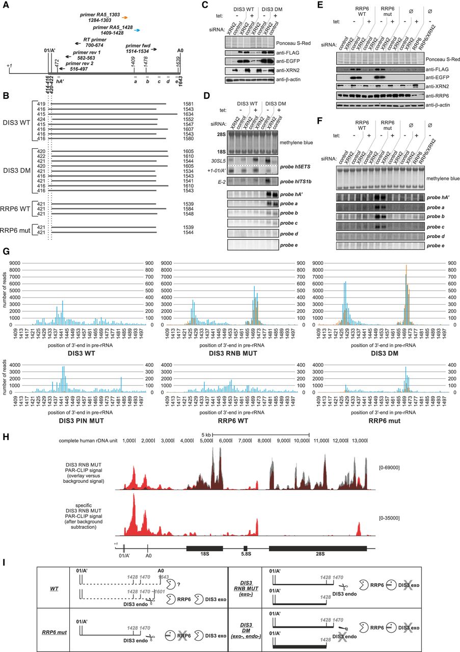
01/A′–A0 processing by-product is degraded in human cells in the 3′–5′ direction by the cooperative action of RRP6 and DIS3, but not by XRN2. (A) Location of primers (arrows) and probes (gray bars) utilized in cRT-PCR (RT, fwd, rev1, rev2), 3′-RACE-seq (RA5_1428, RA5_1303), and northern blot (hA′, a–e) analyses, within 01/A′–A0 fragment of the 5′-ETS. (B) cRT–PCR mapping of 5′-ETS 01/A′–A0 decay intermediates present in model cell lines producing DIS3 WT or DM and RRP6 WT or mut proteins. Each line represents an individual sequenced insert from Topo cloning. Numbers on the left and the right indicate 5′- and 3′-ends of the identified fragments, respectively. Vertical dotted lines show positions of the doublet 01/A′ cleavage site. With one exception, the 5′ terminus of the 01/A′ decay intermediates is intact, indicating that the 5′–3′ exonuclease XRN2 is not involved in the elimination of this species. (C) Model cell lines able to produce exogenous DIS3 WT or DM variants were either untreated (“tet: −”) or treated (“tet: +”) with tetracycline, and transfected with siRNA against XRN2 or with control, unrelated siRNA; proteins extracted from cells were separated in SDS-PAGE and transferred onto nitrocellulose membrane, which was stained with Ponceau S-Red and then sequentially probed with antibodies specific to FLAG epitope, EGFP, XRN2, and β-actin (loading control). (D) Northern blot analysis for RNA samples isolated from model cell lines described in panel C. Total RNA was separated in the denaturing agarose-formaldehyde gel and transferred onto nylon membrane, which was then stained with methylene blue and sequentially hybridized with different probes, as indicated on the right. Analysis of 5′-ETS degradation intermediates with probes hA′ and a–e showed that XRN2 silencing does not exert any additive effect on the accumulation of 01/A′–A0 decay intermediates in cells producing DIS3 DM protein variant. Probes h5ETS and hITS1b were additionally used to demonstrate phenotypes characteristic to XRN2 depletion—positions of RNA species accumulating upon XRN2 down-regulation (30SL5 aberrant precursor and +1-01/A′ and E-2 processing by-products) are indicated on the left. (E) Western blot analysis was performed as in panel C, for protein samples isolated from model cell lines able to produce exogenous RRP6 WT or mut, as well as for parental Hek293 Flp-In T-REx cells (Ø). In addition, the latter cells were subjected to RNA interference using siRNA targeting RRP6 alone or in combination with XRN2 (two rightmost lanes). (F) Northern blot analysis for RNA samples isolated from model cell lines described in panel E. XRN2 down-regulation did not exert any synergistic effect on the accumulation of truncated 01/A′–A0 5′-ETS fragment in conjunction with RRP6 catalytic mutation or siRNA-mediated depletion. (G) 3′-RACE-seq analysis of the 01/A′–A0 decay intermediates in cells producing different DIS3 and RRP6 protein variants. Blue bars and scale on the left of each graph represent the number of reads, the 3′-end of which mapped to a given position in pre-rRNA (indicated at the bottom) in the analysis with 3′-RACE primer RA5_1428; orange bars and scale on the right of each graph correspond to the experiment performed using 3′-RACE primer RA5_1303 (see panel A for the location of 3-RACE primers). (H) Genome Browser screenshots of DIS3 PAR-CLIP data mapped to the consensus sequence of the complete human rDNA repeating unit. Each track represents the mean signal of two biological replicates. Upper part shows the overlay of DIS3 PAR-CLIP signal (red) and the background signal (gray). Lower part represents a specific signal, calculated by subtraction of the background mock signal from the PAR-CLIP signal obtained for DIS3 RNB MUT-expressing cells. Coverage signal corrected to mapped library is shown in size square parentheses on the right of each track. A simplified rRNA precursor scheme is shown at the bottom, with 01/A′ and A0 sites marked with vertical lines. (I) A model for exosome-mediated 01/A′–A0 by-product decay. In wild-type cells, 01/A′–A0 fragment arising after endonucleolytic cleavages is rapidly attacked first by an unknown enzyme and then by the exosome-associated nucleases, and efficiently degraded. RRP6 is mainly involved in the first phase of exosome-mediated decay, but DIS3 also contributes to this process. In turn, further efficient degradation of 01/A′–A0 species requires both catalytic activities of DIS3. Upon their impairment, RRP6 activity is insufficient to eliminate this by-product, which results in a profound accumulation of decay intermediates ending approximately at position 1428 of pre-rRNA.




