


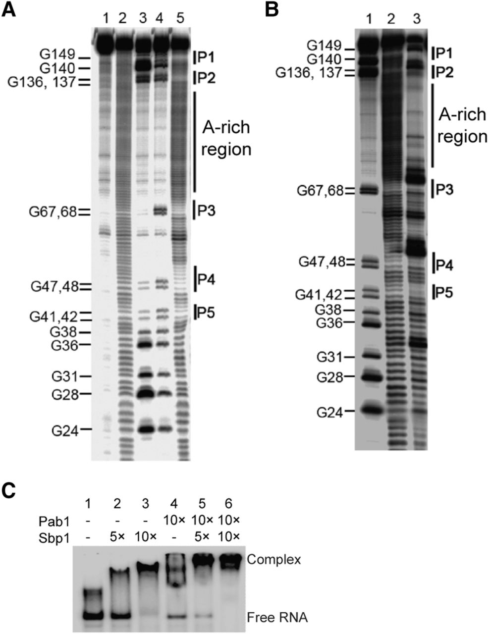
Sbp1 binds to the A-rich region in 5′UTRPab1. (A) An in-line probing gel image of 5′UTRPab1. The 5′UTRPab1 in its undigested form (lane 1) was subjected to partial alkaline digestion (lane 2) and RNase T1 treatment in the presence of urea at 37°C (lane 3) and at 55°C (lane 4). Results on in-line probing of 5′UTRPab1 (lane 5) are shown. Double-stranded regions (P1, P2, P3, P4, and P5) and poly(A)-rich region in the RNA are shown. (B) An in-line probing gel image of 5′UTRPab1 in the presence of Sbp1. 5′UTRPab1 RNA partially digested by RNase T1 at 55°C in the presence of urea (lane 1). The cleavage pattern of 5′UTRPab1 after the in-line probing without Sbp1 (lane 2) and with Sbp1 (lane 3) showing protection of the A-rich region by Sbp1. (C) An EMSA showing the formation of 5′UTRPab1•Sbp1•Pab1 complex. Addition of Sbp1 or Pab1 leads to formation of the RNP complexes of 5′UTRPab1•Sbp1 (lanes 2 and 3) or 5′UTRPab1•Pab1 (lane 4), respectively, and results in a slower electrophoretic mobility of the 32P-labeled 5′UTRPab1. In the presence of Sbp1 and Pab1 simultaneously, a band that migrates even slower appears, suggesting the formation of 5′UTRPab1•Sbp1•Pab1 complex (lanes 5 and 6). Lane 1 shows 5′UTRPab1 only.




