


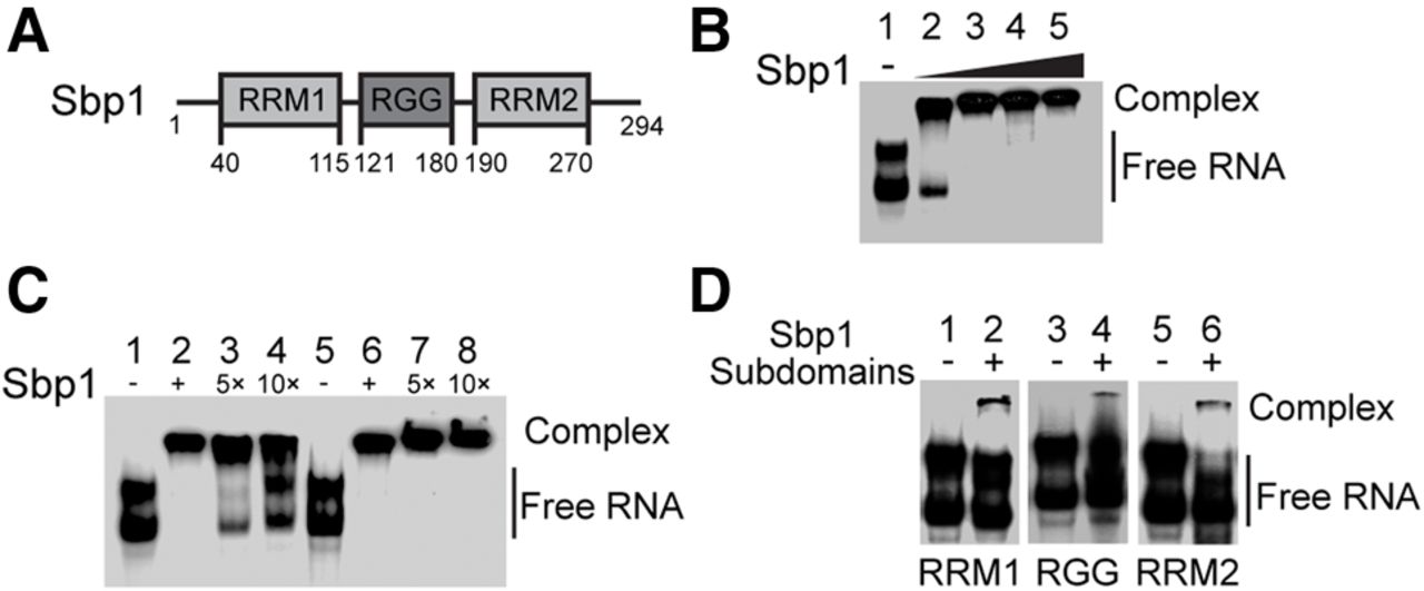
Sbp1 binds to 5′UTRPab1 specifically and cooperatively. (A) Schematic representation of Sbp1's domain organization. Amino acid residue numbers at the boundaries of individual domains are indicated. (B) An EMSA showing the formation of 5′UTRPab1•Sbp1 complex at 1:1 stoichiometry. 5′UTRPab1 was incubated with increasing concentration of purified Sbp1 (1:0, 1:5, 1:10, 1:15, and 1:20 of 5′UTRPab1:Sbp1 for lanes 1–5, respectively). (C) Competition EMSA showing specific interactions between 5′UTRPab1 and Sbp1. Sbp1 was first incubated with 32P-5′UTRPab1 (lanes 2 and 6). An excess of unlabeled 5′UTRPab1 by five- and 10-fold (lanes 3 and 4) and its complementary RNA, 5′UTRPab1-complement, (lanes 7 and 8) were used to compete with 32P-5′UTRPab1 bound to Sbp1. (D) An EMSA showing weak interactions between the 5′UTRPab1 and individual domains of Sbp1. Sbp1RRM1 (lane 2), Sbp1RGG (lane 4), and Sbp1RRM2 (lane 6) were incubated with 5′UTRPab1. A molar ratio of 1:20 (5′UTRPab1:Sbp1 domain mutants) was used in the experiment. 5′UTRPab1 alone was shown in lanes 1, 3, and 5.




