


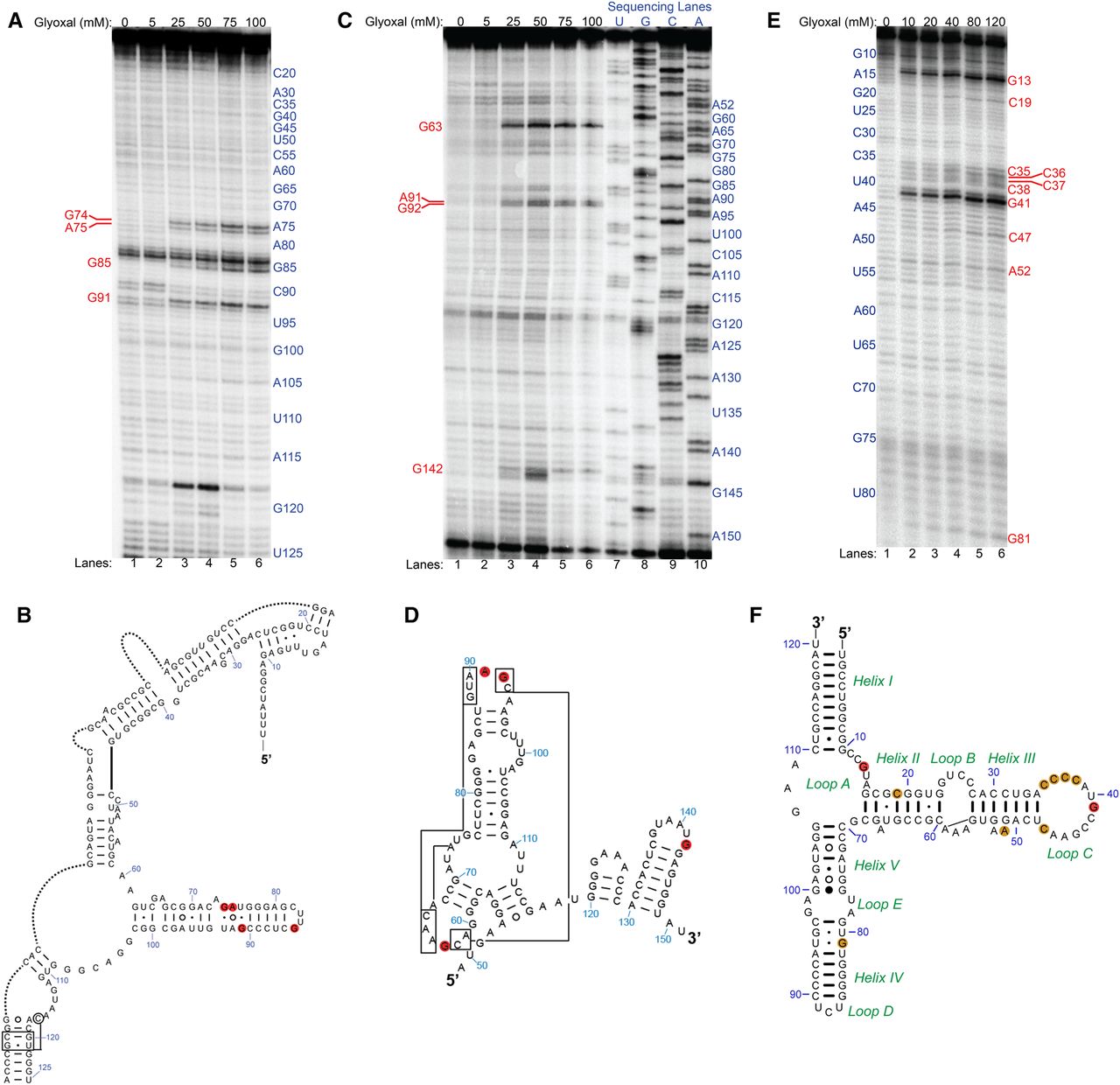
In vivo glyoxal modification of B. subtilis and E. coli rRNAs. (A) Denaturing PAGE analysis of cDNAs generated after reverse transcription (RT) for B. subtilis 16S rRNA. Shown are reactions in glyoxal from 0 to 100 mM. Reactive nucleotides are shown in red text. (B) Reactive nucleotides mapped as red discs onto the relevant portion of B. subtilis 16S rRNA comparative secondary structure. Portions of the structure not relevant to this study were removed and replaced with dotted lines. (C) Denaturing PAGE after RT for B. subtilis 23S rRNA. Shown are reactions in glyoxal from 0 mM to 100 mM and dideoxy sequencing lanes. Reactive nucleotides are shown in red text. (D) Reactive nucleotides mapped as red discs onto the relevant portion of B. subtilis 23S rRNA comparative secondary structure. (E) Denaturing PAGE after RT for E. coli 5S rRNA. Shown are reactions in glyoxal from 0 to 120 mM. Reactive nucleotides are shown in red text. (F) Reactive nucleotides mapped as orange and red discs onto the E. coli 5S rRNA comparative secondary structure. Orange discs indicate moderate glyoxalation of a given nucleotide while red discs indicate strong glyoxalation at that nucleotide.




