


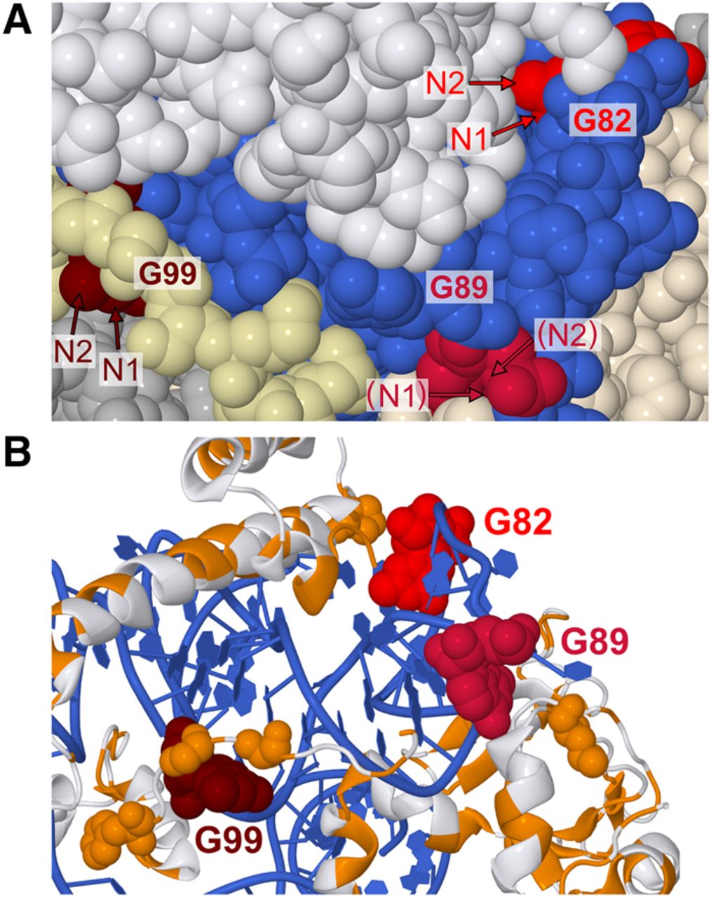
Cryo-EM structure of Saccharomyces cerevisiae 60S subunit (PDB: 5GAK), a homolog of rice 60S subunit used here as no rice ribosome structure currently exists. (A) Relative orientations of N1 and N2, the atoms responsible for glyoxal reactivity. 5.8S rRNA is blue, G82 is scarlet, G89 is crimson, and G99 is maroon. 28S rRNA and other RNAs are silver. Ribosomal proteins are white, except for two ribosomal proteins in the immediate vicinity of G89 and G99, which are tan (L26-A) and pale yellow (L37-A). Arrows indicate N1 and N2 atoms, with parentheses indicating N1 or N2 obscured by other atoms. (B) Proximity of G82, G89, and G99 to hydrophobic protein residues. Only 5.8S rRNA and ribosomal proteins L26-A, L35-A, L37-A, are shown. Hydrophobic residues are orange and nonhydrophobic residues are white while G82, G89, and G99 are colored as in panels A and B. Hydrophobic residues within 10 Å of N1 or N2 atoms in reactive guanines are shown as spheres.




