


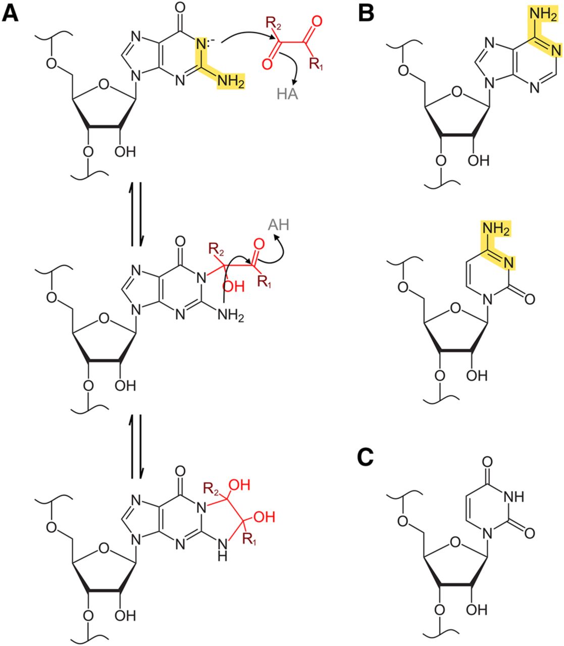
Reaction scheme for base glyoxalation by glyoxal and derivative reagents. (A) Reaction of a generalized glyoxal reagent with guanosine. R1 is H in glyoxal, CH3 in methylglyoxal, C6H5 in phenylglyoxal, and CH3 in dimethylglyoxal. R2 is H in glyoxal, methylglyoxal, and phenylglyoxal, and is CH3 in dimethylglyoxal. Highlighted in yellow is the amidine moiety of the guanine base, which is required for the glyoxalation reaction and is also present in adenine and cytosine bases but absent in uracil. In the first reaction step, deprotonated N1 attacks glyoxal, methylglyoxal, or phenylglyoxal at the more electronegative formyl carbon. In the following step, N2 then attacks at the keto carbon, leading to formation of a cyclic adduct between N1 and N2. In the case of dimethylglyoxal reactions, both carbons are keto carbons, but the generalized scheme is the same. (B) Adenine and cytosine also possess an amidine moiety, highlighted in yellow. (C) Uracil lacks an amidine moiety, which prevents its reactivity with glyoxal or similar reagents.




