


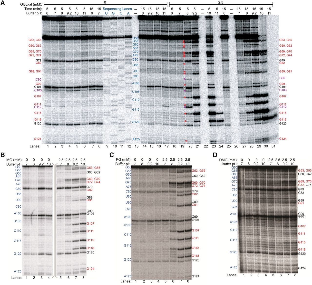
In vitro modification of rice 5.8S rRNA by glyoxal and derivatives analyzed by denaturing PAGE of cDNAs after reverse transcription. (A) Glyoxal reactions. Control reactions, reactions with 2.5 mM glyoxal, and dideoxy sequencing lanes are shown. Lanes 13, 22, and 26 were not loaded. Positions of all guanines within the examined range (G53 to G124) are marked on the sides of the gel with text, or between lanes 19 and 20 with asterisks, with red denoting glyoxalated guanines, violet denoting glyoxalated cytosines at pH 8 and 5 min time, and black denoting natural reverse transcriptase stops. (B) Methylglyoxal reactions. (C) Phenylglyoxal reactions. (D) Dimethylglyoxal reactions. In panels B–D, reactive guanines are shown in red text, unreactive guanines in black text, and natural reverse transcription stops in outlined text.




