


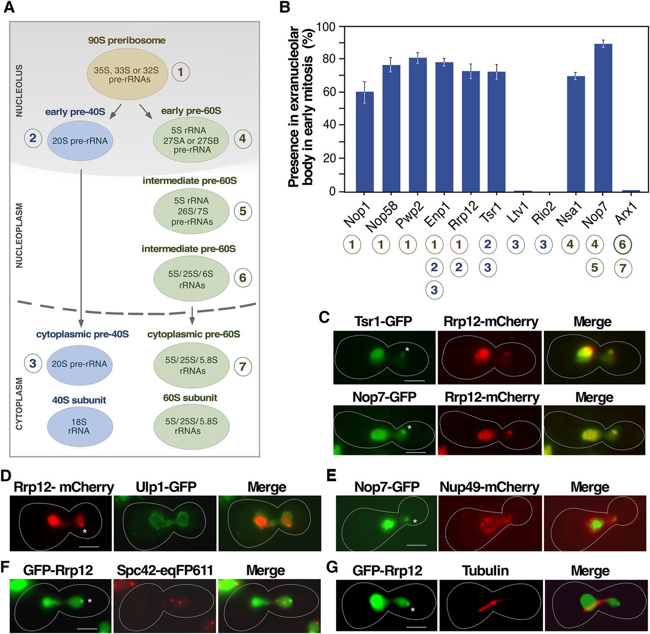
The Tsr1+ extranucleolar body contains both 40S and 60S preribosome particle components. (A) Scheme of the maturation of ribosomal subunits in S. cerevisiae. (B) Quantification of the presence in the extranucleolar body of the indicated GFP-tagged proteins in early mitosis cells. Circled numbers (bottom) indicate the specific association of those proteins with the preribosomal particles depicted in panel A. (C–F) Representative images of indicated GFP- (green color), mCherry- (red color), and eqFP611- (red color) tagged proteins in cells transiting early mitosis. (G) Representative image of endogenous tubulin (red color) detected by standard immunofluorescence techniques in cells expressing GFP-Rrp12 (green color). In panels B–G, the experiments were performed with cells 40 min upon release from hydroxyurea arrest following the scheme outlined in Figure 1A. In panels C–G, areas of colocalization are shown in yellow. Asterisks indicate the position of the extranucleolar preribosome body. Scale bars, 2 µm.




