


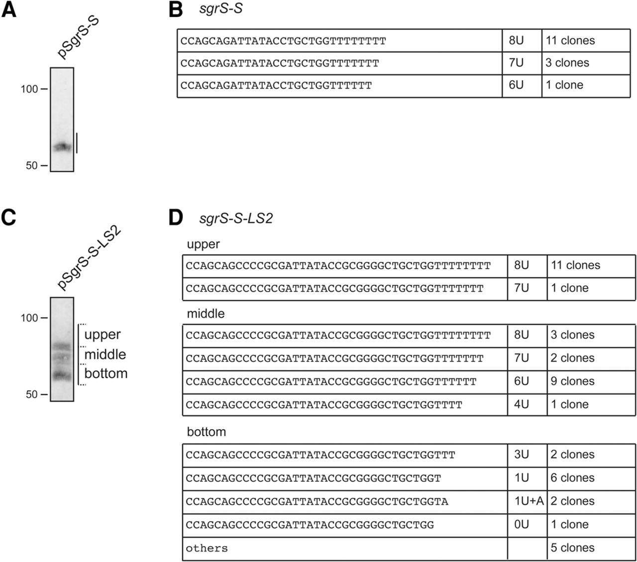
Analysis of the 3′ ends of transcripts derived from sgrS-S and sgrS-S-LS2. TM542 (ΔsgrS) cells harboring pSgrS-S (A) or pSgrS-S-LS2 (C) were grown in LB medium. At A600 = 0.6, 0.1% αMG was added and incubation was continued for 10 min, and then 0.02% arabinose was added and incubation was continued for 5 min. Total RNAs were prepared and duplicate RNA samples (10 µg) were resolved side-by-side on a 12% polyacrylamide gel electrophoresis in the presence of 8 M urea. One side of the gel was subjected to Northern blotting (A,C). The region corresponding to transcripts was cut out from the other side of the gel (A,C). A single gel piece containing the SgrS-S band was used for transcripts from sgrS-S (A). The gel was divided into three gel pieces (upper, middle, and lower) for transcripts from sgrS-S-LS2 (C). The upper gel piece corresponds to the full-length transcript, whereas the middle and lower gel pieces correspond to heterogeneous shorter transcripts. RNAs were purified from the gel pieces and subjected to 3′-RACE analysis. DNA sequences corresponding to the 3′ region of transcripts were determined by using randomly picked-up plasmid clones containing amplified cDNAs. The sequences and number of clones analyzed are shown (B,D). Others represent the clones in which the 3′ ends of RNAs were mapped in the region prior to the terminator T residue stretch (D).




