


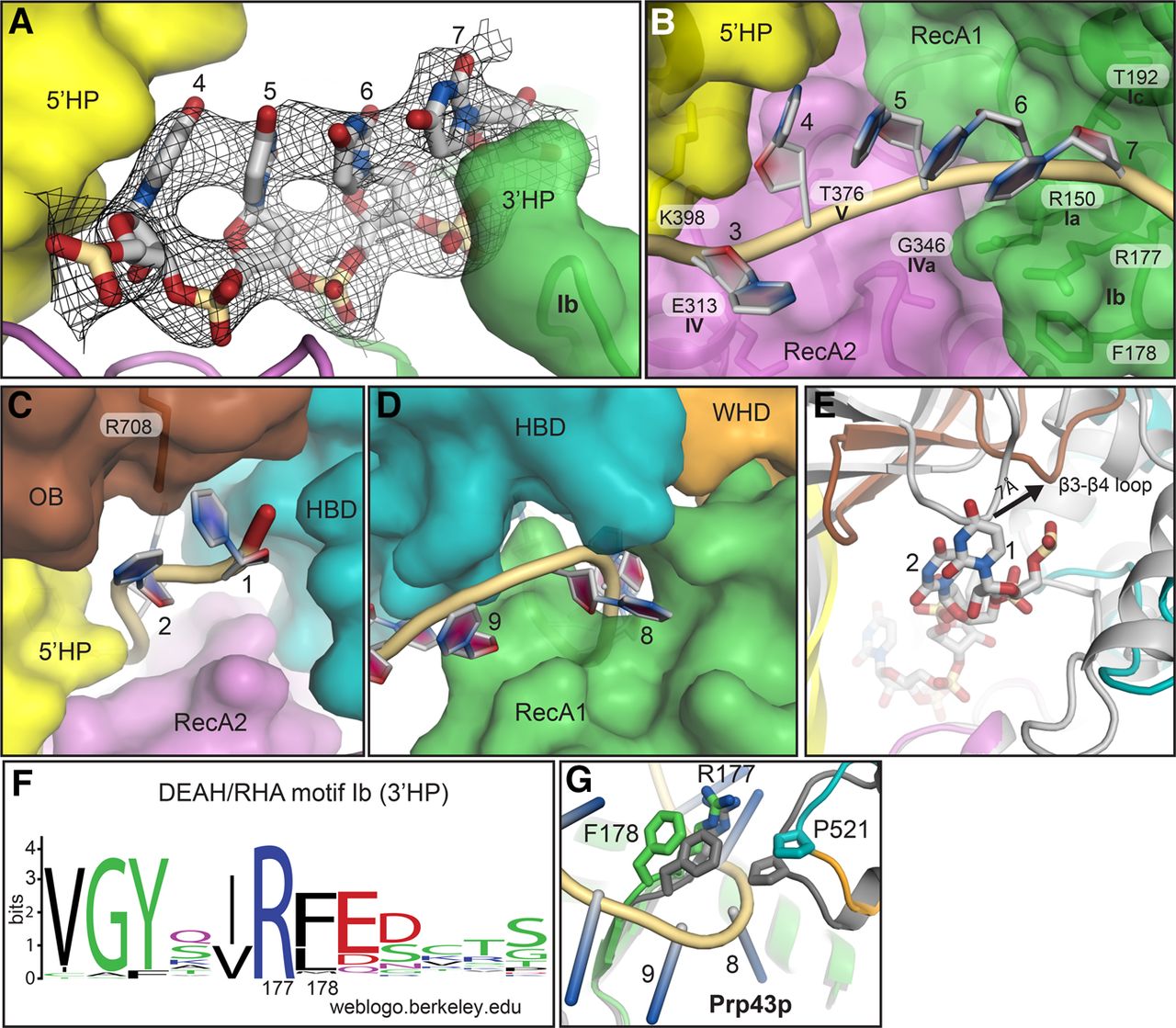
RNA binding by Prp43p involving motif Ib. (A) A 2mFo–DFc electron density omit map surrounding four stacked RNA bases is shown, averaged and contoured at 1.0 σ. For clarity, only part of the RecA1 and RecA2 domains are shown with surface representation of the 5′HP and motif Ib. (B–D) Cartoon representation of RNA and surface representation of domains, demonstrating proximity of conserved motifs and bases, as indicated. (B) Central four stacked bases. (C) Entry channel. (D) Exit channel. (E) A comparison of Prp43p in the ADP-bound (PDB 3KX2) (gray) and the ADPNP–RNA-bound (colored) states, with the RecA2 domains superimposed to reveal movement of the OB fold required to accommodate RNA. (F) The RNA-binding RF dipeptide at the tip of the 3′HP, encompassing motif Ib, is conserved. The sequence logo shows conservation within the 3′HP among human, Drosophila melanogaster, Arabidopsis thaliana, and yeast DEAH/RHA ATPases (see Materials and Methods for sequences). (G) RNA binding is accommodated by opening of the RNA exit channel by separation of the 3′HP and a proline of the auxiliary domain. The closed RNA-free, ADP-bound structure is shown in gray; the open in RNA–ADPNP-bound structure is colored as in Figure 1.




