


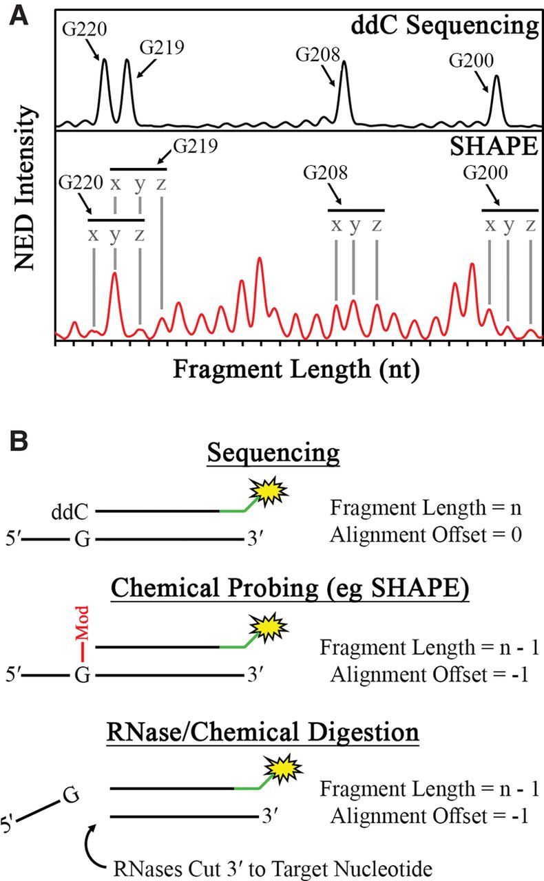
Schematic description of sequence alignment in RiboCAT. (A) To optimize the assignment of peaks to their corresponding nucleotides in the RNA sequence, an initial guess of the alignment is made (peaks labeled “y”) based on the x-axis similarities in peaks from the sequencing (top) and experimental (bottom) traces. The alignment is then shifted incrementally to the left (peaks labeled “x”) or incrementally to the right (peaks labeled “z”), and the RMSD between the x-axis values are calculated. The minimum RMSD over the entire trace is chosen as the correct alignment. (B) In standard Sanger sequencing, the DNA fragment corresponding to a particular residue will include the cognate dideoxy nucleotide (top); however, in both chemical probing methods (middle) and RNase/chemical digestion (bottom), the cognate nucleotide is prohibited from being incorporated by either a chemical modification or backbone cleavage, respectively, leading to an offset of −1.




