


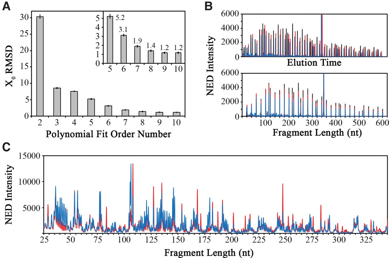
FIGURE 2.
Signal alignment using an internal size standard. (A) Optimization of the polynomial order used for signal alignment. The inset shows a zoomed region encompassing orders 5–10 with RMSD values reaching a plateau at an average of X0 = 1.2 at order 9. (B) The unaligned (top) and aligned (bottom) size standards for three separate CE experiments of sequencing (black), no reagent control (blue), and SHAPE reaction (red). (C) Aligned SHAPE reaction traces for no reagent control (blue) and SHAPE reagent (red) show highly accurate signal alignment based on the internal size standard.




