


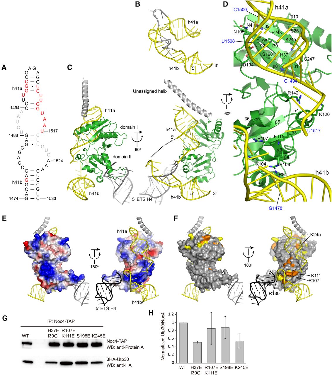
Interaction of Utp30 with 90S. (A) Secondary structure of 18S rRNA helix 41. The nucleotides contacted by Utp30 are colored in red and unmodeled in 90S are in gray. The top and base stems of helix 41 are termed as 41a and 41b. (B) Structure of helix 41 in mature 40S ribosome. The h41b structure is aligned to its counterpart in the right panel of C. The nucleotides unmodeled in 90S are colored in gray. (C) Interactions of Utp30 with 90S. (D) Detailed interactions of Utp30 with helix 41. Residues at the RNA-binding interface are shown as sticks. (E) Electrostatic potential surface of Utp30. The surface is colored from blue to red for positively to negatively charged regions. (F) Conservation surface of Utp30. The residues that are conserved in 100% and 80% of nine aligned Utp30/Cic1 sequences in Figure 1C are colored in orange and yellow, respectively. (G) Association of mutant Utp30 to 90S. The utp30Δ/Noc4-TAP strain was transformed with a pRS415 plasmid expressing wild-type (WT) or mutant Utp30. The Noc4-TAP particle was immunoprecipitated (IP) with IgG-coated beads and analyzed with SDS-PAGE and Western blotting. (H) Quantification of Utp30 binding. The Utp30 to Noc4 volume ratio was calculated and normalized against WT Utp30. Averages of three experiments and standard deviations are shown.




