


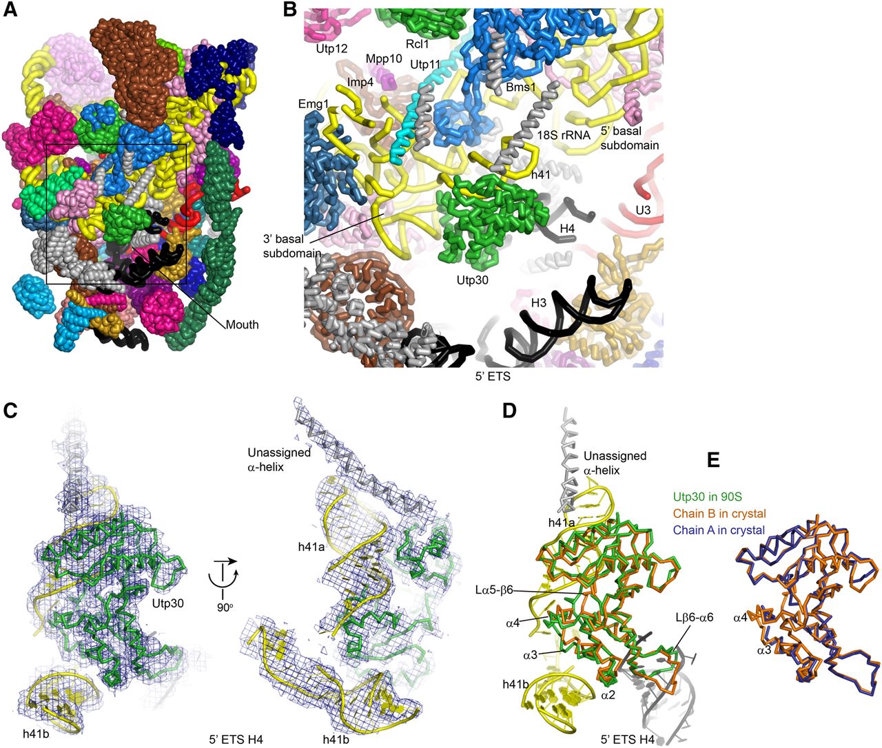
FIGURE 2.
Structure of Utp30 in 90S. (A) Structure of 90S preribosome showing the front face. Individual proteins are color-coded and unassigned proteins are in gray. The 18S, 5′ ETS, and U3 RNAs are colored yellow, black, and red, respectively. (B) Zoomed-in view of the mouth. (C) Cryo-EM density map of the ΔMtr4 90S is fitted with Utp30 and its binding partners. Two perpendicular views are shown. (D) Alignment of Utp30 structures in crystal (orange) and in 90S (green). (E) Structural alignment of two Utp30 protomers in crystal.




