


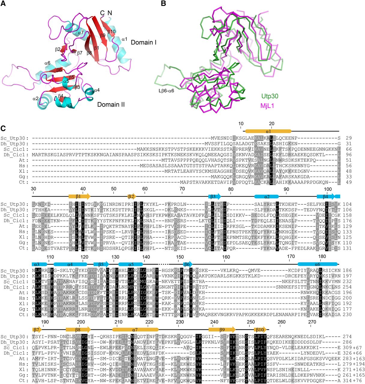
Crystal structure of Utp30. (A) Ribbon representation of Utp30 structure. The α-helices, β-strands, and loops are colored in cyan, red, and magenta, respectively. The secondary structures, N and C termini, and two domains are labeled. (B) Structural alignment between Utp30 and L1 from Methanococcus jannaschii (MjL1, PDB code 1U63). (C) Multiple sequence alignment of Utp30/Cic1 family proteins. The sequences are from Saccharomyces cerevisiae (Sc), Debaryomyces hansenii (Dh), Arabidopsis thaliana (At), Homo sapiens (Hs), Xenopus laevis (Xl), Gallus gallus (Gg), and Chaetomium thermophilum (Ct). Residues with 100%, 80%, and 60% conservation are shaded in black, gray, and light gray, respectively. The secondary structures observed in the crystal structure of Utp30 are shown on the top and colored by domain.




