


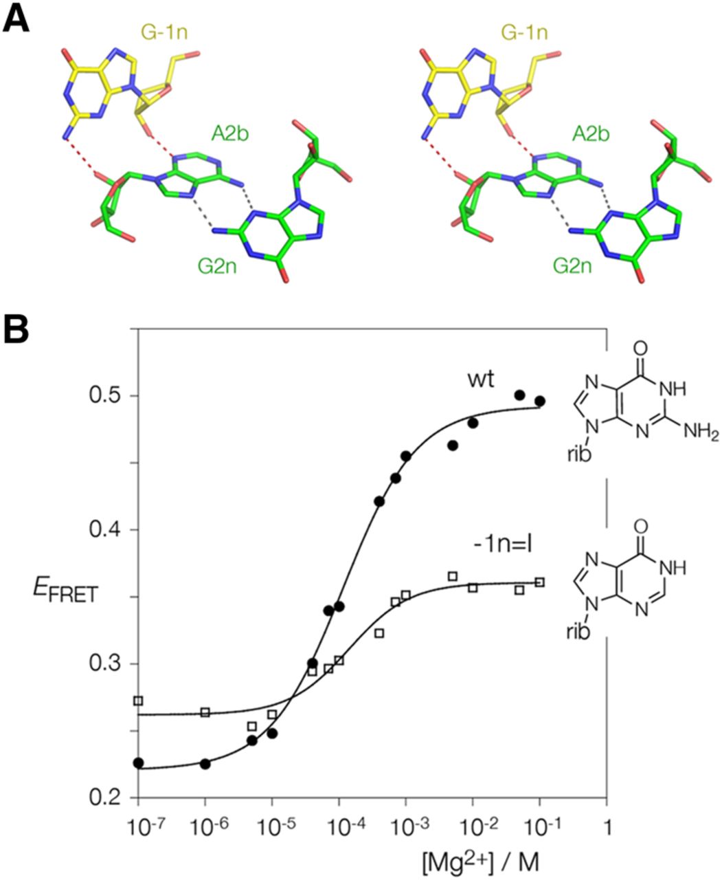
Atomic mutagenesis to probe the importance of the hydrogen bonding between G-1n and A2b. (A) Parallel-eye stereoscopic image of the interaction of G-1n with A2b in Hm Kt-7. The cross-strand hydrogen bonds are colored red. Note the additional hydrogen bond from G-1n N2 to the O2′ of A2b. This image was prepared using PDB ID 4CS1. (B) A modified version of the Kt-7 construct used for FRET analysis was prepared in which G-1n was replaced by inosine, thus replacing the amine at the 2 position with a proton. Folding as a function of Mg2+ ion concentration was studied by measurement of FRET efficiency between the terminally attached fluorophores as before. The data are plotted for unmodified Kt-7 (filled circles) and the G-1nI variant, and fitted to the two-state folding model. Note that the folding of the variant (open squares) lacking the G-1n N2 is significantly impaired.




