


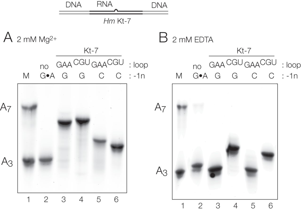
Analysis of ion-dependent folding of H. marismortui Kt-7 k-turn using gel electrophoresis. The schematic (top) shows the constructs used in these studies. The k-turn is centrally located in an RNA section embedded within a longer DNA segment giving a total length of 65 bp excluding the bulge. The different species were electrophoresed in a 13% polyacrylamide gel in the presence of (A) 2 mM Mg2+ ions or (B) 2 mM EDTA. To provide a frame of reference, equivalent duplexes in which the k-turn was replaced by an A3 or A7 bulge were mixed and electrophoresed in track 1. Track 2 contains a Kt-7 variant where the three G•A base pairs have been changed to Watson–Crick G–C base pairs by the substitutions of A1n, A2b, and A3b to C. Tracks 3 to 6 contain Hm Kt-7 variants with various substitutions taken from Af box C/D k-turn. Track 3, unmodified Kt-7; track 4 loop changed to CGU; track 5, −1b:−1n = G:C; track 4, −1b:–1n = G:C plus loop = CGU.




