


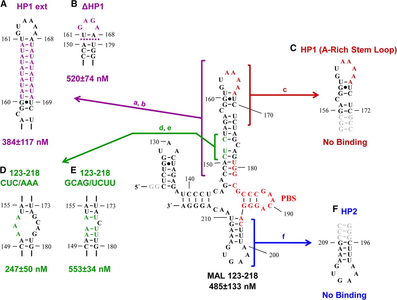
FIGURE 2.
Predicted secondary structure of MAL 123–218 (Goldschmidt et al. 2004). The PBS is highlighted in red, and the two 5′ G's shown in gray were added to this construct to facilitate in vitro transcription. (A–F) The various stem–loops and mutations used in the binding assays are indicated, as described in the text. In the case of HP1 and HP2, three additional closing G–C base pairs were added to stabilize the stems (gray). The apparent dissociation constants for the binding of each RNA construct to hLysRSΔN65 are indicated.




