


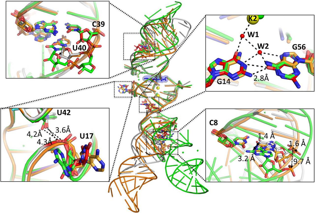
FIGURE 4.
Superposition of different Spinach/DFHBI complex crystal structures. The structure obtained in the present study is shown in gray and is superimposed to that of Spinach-FdA (shown in green) and that of Spinach-P (shown in orange). The regions containing the beneficial mutations are zoomed-in and the mutated residues are shown in red, whereas the wild-type ones are shown according to the color code used before. Potassium ions are represented by yellow spheres and water molecules by red dots.




