


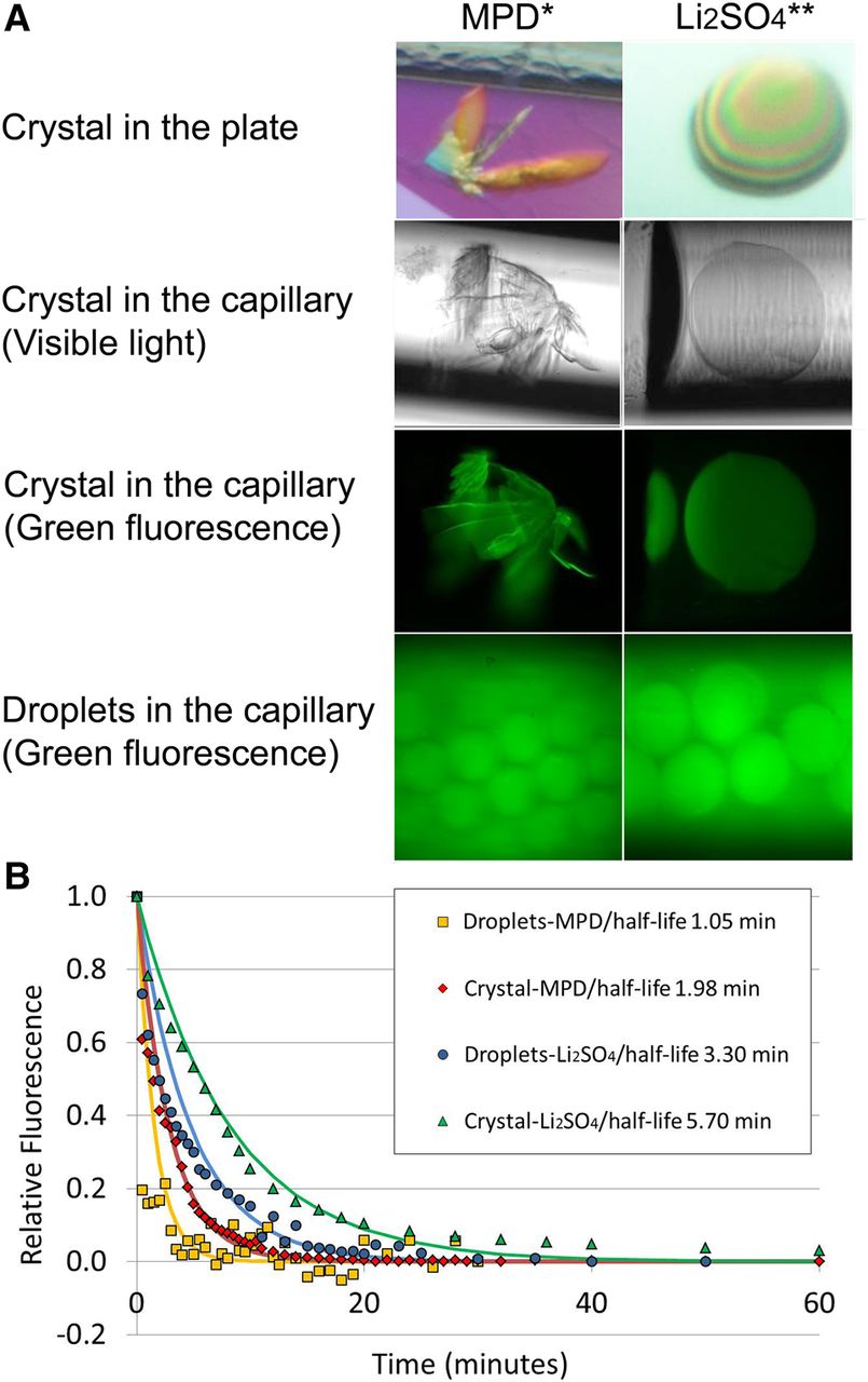
Fluorescence properties of the iSpinach-D5-G30–A32/DFHBI crystallized complex. (A) Micrographs of crystals and droplets. Micrographs of the crystals were taken while they were in the crystallization plate or loaded into the capillary and observed under visible light or in epi-fluorescence mode. The conditions in which the crystal was grown or the droplets prepared (MPD or Li2SO4) are indicated under the corresponding column. (*) MPD crystallization conditions were 32% MPD, 40 mM NaCl, 40 mM sodium cacodylate pH 7.0 and 12 mM spermine. (**) Li2SO4 crystallization conditions were 1.9 M Li2SO4, 50 mM MES pH 5.6, 5 mM MgCl2. (B) Fluorescence decay monitoring. The fluorescence of the crystals or the droplets was monitored along the time. The values were relativized to that of the first measurement point, and an exponential decay was fit to the data to compute the fluorescence half-life (values given in the legend of the plot).




