


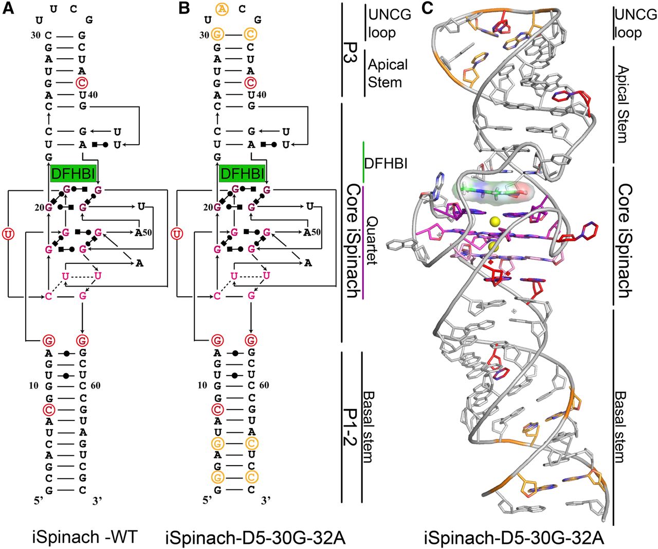
FIGURE 1.
Structural organization of iSpinach and its crystallized mutant. (A) Secondary structure of iSpinach. (B) Secondary structure of iSpinach-D5-G30–A32. On both models, the beneficial mutations combined to obtain iSpinach are shown and circled in red, while those introduced to increase the transcription rate of the molecule and promote its crystallization are shown in orange. (C) Crystal structure of iSpinach-D5-G30–A32. The residues are shown using the same color code used on secondary structure models. Potassium ions are shown as yellow spheres.




