


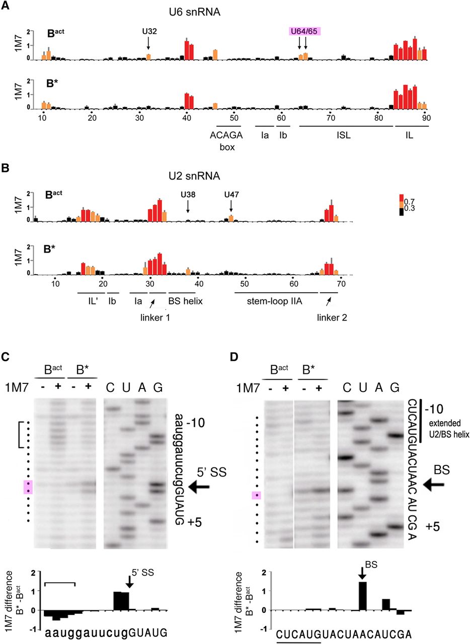
SHAPE analysis of the RNA–RNA network in yeast Bact and B* spliceosomes. (A,B) Shape analysis of U6 snRNA (A) and U2 snRNA (B) in Bact and B* complexes, as indicated. The data represent the mean of three independent experiments. Normalized reactivities are color-coded (inset) into three different categories of flexibility as previously described (Mortimer and Weeks 2007): black (0–0.3; not flexible, helically constrained), orange (0.3–0.7; intermediate flexibility, no internal Watson/Crick base pairs), and red (>0.7; fully unconstrained, single stranded). Key nucleotides are labeled above the top graph in each panel. Nucleotide numbers and the positions of structural elements are shown below the bottom graph in each panel. (C,D) Representative gels of the 1M7 reactivities of the pre-mRNA in Bact and B* around the 5′ splice site (C) and the branch site sequence (D) analyzed by primer extension. Primer extension will stop 1 nt before the 2′-O-acylated nucleotide relative to the dideoxy sequence. The bracket in C denotes the stretch of nucleotides with reduced 2′-OH flexibility in the B* complex. Quantifications of the autoradiographs in C and D were used for the difference plots (B* − Bact) shown below the respective autoradiographs. The sequences shown below the difference plots correspond to the actual nucleotide involved in attack or protection from 2′-O-acylation by 1M7.




