


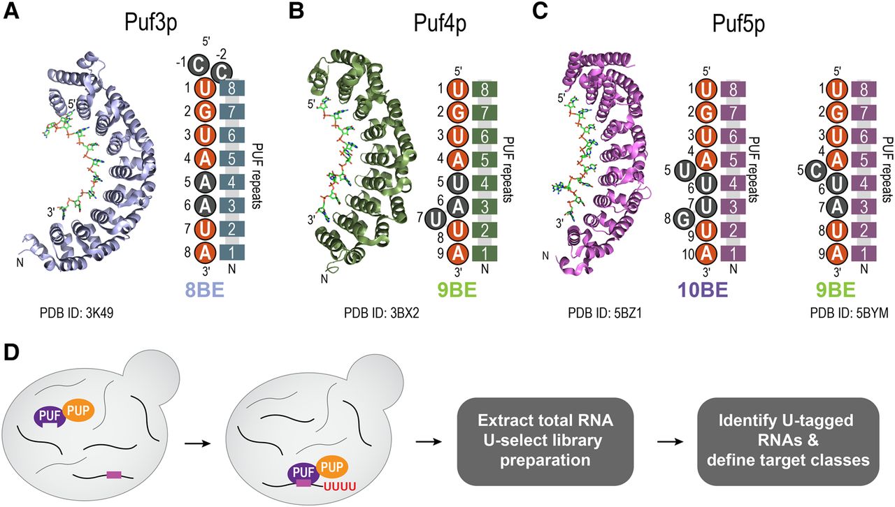
Puf3p, Puf4p, and Puf5p share similar structures and bind distinct yet related RNA sequences. (A) Crystal structure of Puf3p bound to RNA (PDB ID: 3K49) (Zhu et al. 2009), and a cartoon schematic of the PUF domains of Puf3p bound to an 8BE, with upstream cytosines. (B) Crystal structure of Puf4p bound to RNA (PDB ID: 3BX2) (Miller et al. 2008), and a cartoon schematic of the PUF domains of Puf4p bound to a 9BE. (C) Crystal structure of Puf5p bound to RNA (PDB ID: 5BZ1) (Wilinski et al. 2015), and a cartoon schematic of the PUF domains of Puf5p bound to a 10BE and a 9BE (based off PDB ID: 5BYM). In panels A–C, the RNA-binding domains of Puf3p, Puf4p, and Puf5p, which contain the 8 PUF repeats, are represented by the eight connected rectangles. The numbers in the rectangles indicate the PUF repeat identity, numbered from N to C terminus. The nucleotides represented by orange circles represent the common sequence typically shared by an 8BE, 9BE, and 10BE. The nucleotides in gray represent the regions that distinguish the binding elements. (D) Schematic of RNA Tagging. PUP-2 from C. elegans was fused to the C terminus of the endogenous copy of Puf3p, Puf4p, and Puf5p. Inside live yeast, the PUF–PUP fusion proteins bind to target mRNAs and deposit a covalent “U-tag” on the 3′ end of the mRNA. After extraction of total RNA under denaturing conditions, U-tagged mRNAs are preferentially enriched in the U-select library preparation and analyzed via paired-end high-throughput sequencing. U-tagged RNAs are computationally identified and analyzed to define mRNA targets and target classes.




