


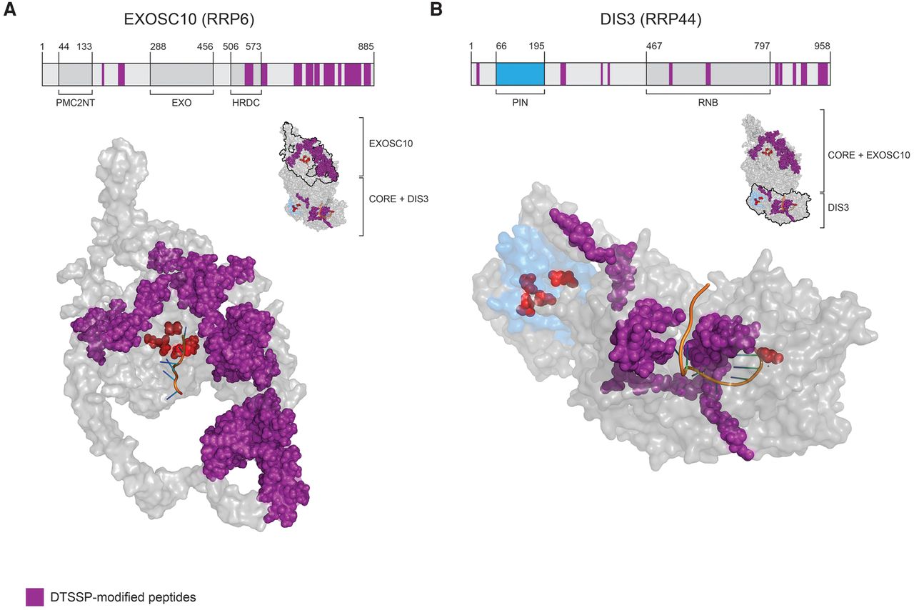
DTSSP-modified peptides mapped to EXOSC10 and DIS3 protein sequences and structures. (A, upper) Linear arrangement of EXOSC10 domains with DTSSP-modified peptides mapped (in purple). The domain organization is based on the protein sequences obtained from the uniprot.org and pfam.xfam.org databases. (Lower) The in silico modeled structure of EXOSC10 using I-TASSER (Yang and Zhang 2015; Yang et al. 2015). Highlighted are DTSSP-modified peptides (purple), key active site residues (red), and cocrystalized RNA (orange). The protein orientation is indicated by the miniature structure of a 12-component exosome (PDB ID: 5c0w with EXOSC10 model), upper right. (B, upper) Arrangement of DIS3 domains represented as in A. (Lower) DTSSP-modified peptides were first mapped onto the human DIS3 protein sequence and the equivalent residues were then mapped on the yeast DIS3 structure (PDB ID: 5c0w), displayed, and colored as in A. Additionally, the PIN domain was labeled in blue to distinguish it from the RNB domain. The protein orientation is indicated as in A.




