


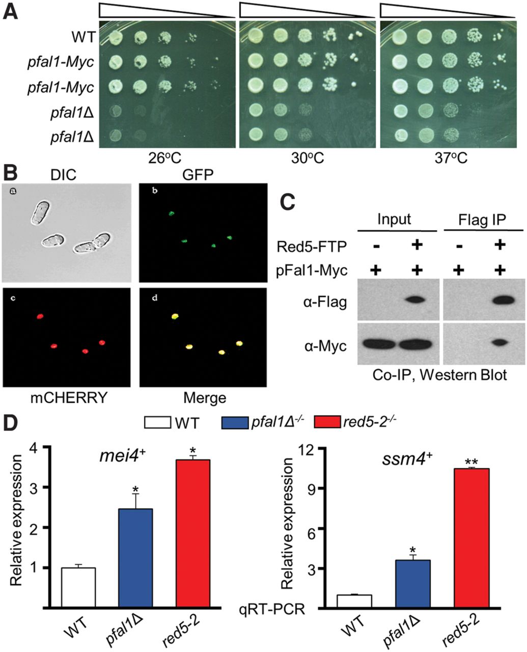
pFal1 localizes to a chromatin-rich region in the nucleus and interacts with Red5. (A) pfal1Δ haploid cells have a severe growth defect. Cells were serially diluted 10-fold, plated on rich media (YEA), and incubated at the indicated temperatures for 3 d. (B) pFal1-GFP colocalizes with Hta1-mCherry in confocal images of live pombe cells. (C) pFal1 interacts with Red5 as determined by Flag Co-IP and Western blot using antibodies as indicated. Immunoprecipitated samples were treated with 20 µg of RNase A/H mixture and 33 µg of DNase I before the final wash. (D) qRT-PCR analysis shows increased mei4+ and ssm4+ transcripts relative to act1+ normalized to wild type (WT = 1), during vegetative growth in diploid cells containing red5-2 or pfal1Δ. Asterisks denote a significant difference comparing each sample with wild type, (*) P ≤ 0.05 and (**) P ≤ 0.005.




