


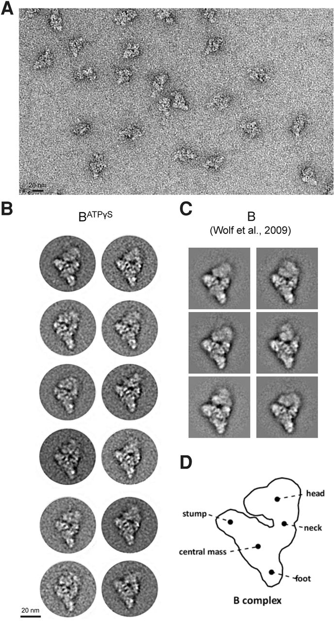
FIGURE 4.
Electron microscopy of affinity-purified BATPγS complexes. (A) Overview of negatively stained BATPγS complexes. (B) Gallery of class averages of BATPγS complexes. (C) Typical class averages of B complexes purified after 6 min of splicing in the presence of ATP (Wolf et al. 2009). (D) Schematic representation of the human B complex, with main structural features labeled.




