


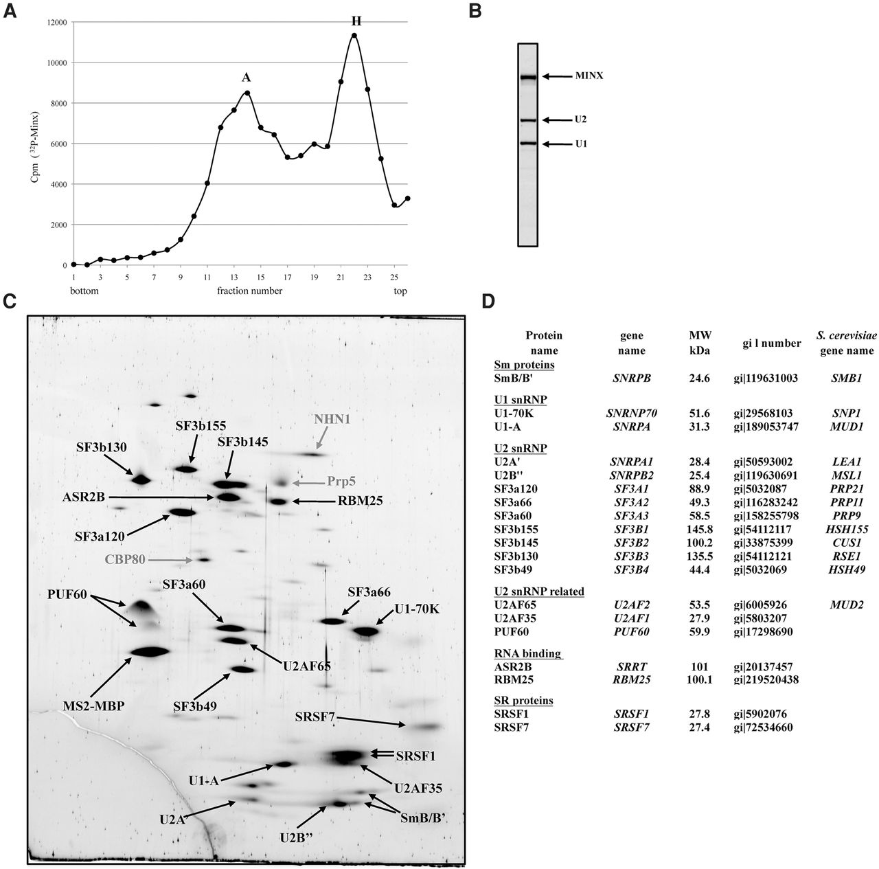
Affinity purification of A complexes accumulating in the presence of ATPγS. (A) Splicing complexes were allowed to form on 32P-MINX pre-mRNA containing MS2 aptamers bound by MS2-MBP protein, in HeLa nuclear extract containing 0.4 mM ATPγS. Complexes were subsequently affinity purified on an amylose column and the eluate subjected to glycerol gradient centrifugation in buffer containing 150 mM NaCl. The number of cpm in each gradient fraction is plotted. (B) RNA composition of complexes in the faster migrating peak (fraction #13). (C) Proteins from affinity-purified A complexes formed in the presence of ATPγS (fractions #13–14) were separated by 2D gel electrophoresis, stained with RuBPS, and the identities of protein spots were determined by mass spectrometry. Proteins smaller than 25 kDa were not analyzed. Abundant proteins are indicated in black and less abundant ones in gray. (D) Summary of abundant/moderately abundant proteins identified in AATPγS complexes.




