


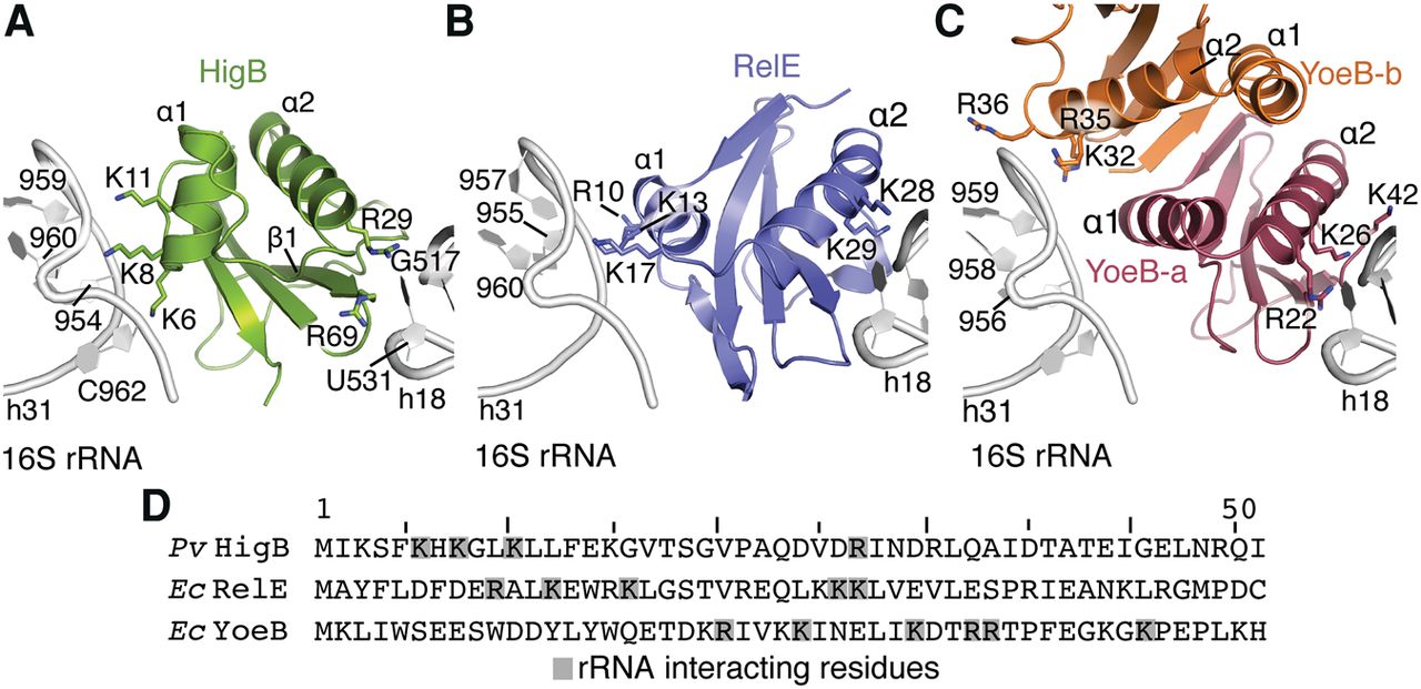
FIGURE 5.
Ribosome-dependent toxins recognize the head domain of the 30S subunit. Views of HigB (A; this study), RelE (B; PDB code 4V7J), and YoeB (C; PDB code 4V8X) interacting with h18 and h31 rRNA via basic residues. The RelE and YoeB structures were captured in the precleavage state and basic residues within hydrogen bonding distance of h18 and h31 are shown as sticks. (D) Alignment of regions of ribosome-dependent toxins Proteus vulgaris (Pv) HigB, E. coli (Ec) RelE, and E. coli (Ec) YoeB that interact with 16S rRNA, demonstrating the low sequence identities.




