


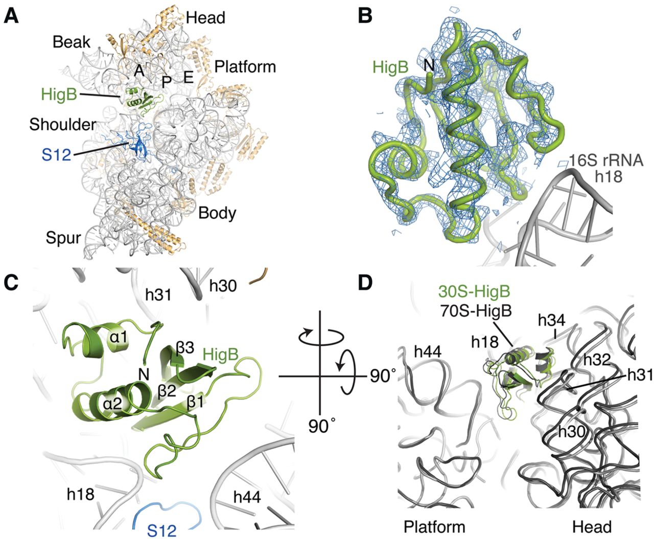
Structural insights into how HigB recognizes the 30S subunit. (A) The X-ray crystal structure of HigB bound to the A site of the 30S subunit. 16S rRNA and ribosomal proteins are shown in gray and tan, respectively, and the aminoacyl (A), peptidyl (P), exit (E) sites, and 30S domains are labeled. (B) Unbiased Fobs–Fobs difference electron density map of the 3.6 Å X-ray crystal structure of HigB bound to the Thermus thermophilus 30S subunit contoured to 1.5σ. Fobs–Fobs difference electron density map was calculated using the structure factors of the apo 30S structure (PDB code 1J5E). (C) Zoomed in view in the same orientation as in A, emphasizing how HigB interacts with multiple 16S rRNA helices and ribosomal protein S12. (D) Comparison of how HigB interacts with the 30S small subunit in the context of the 30S-HigB structure (green; this study) and 70S-HigB ΔH92 precleavage state structure (dark gray; PDB code 4YPB). The 16S rRNA body domain (nucleotides 560–912) of each structure was aligned by least-squares fit in the program Coot (Emsley et al. 2010). 16S rRNA nucleotides from the 30S-HigB and 70S-HigB structures are depicted in light gray and dark gray, respectively.




