


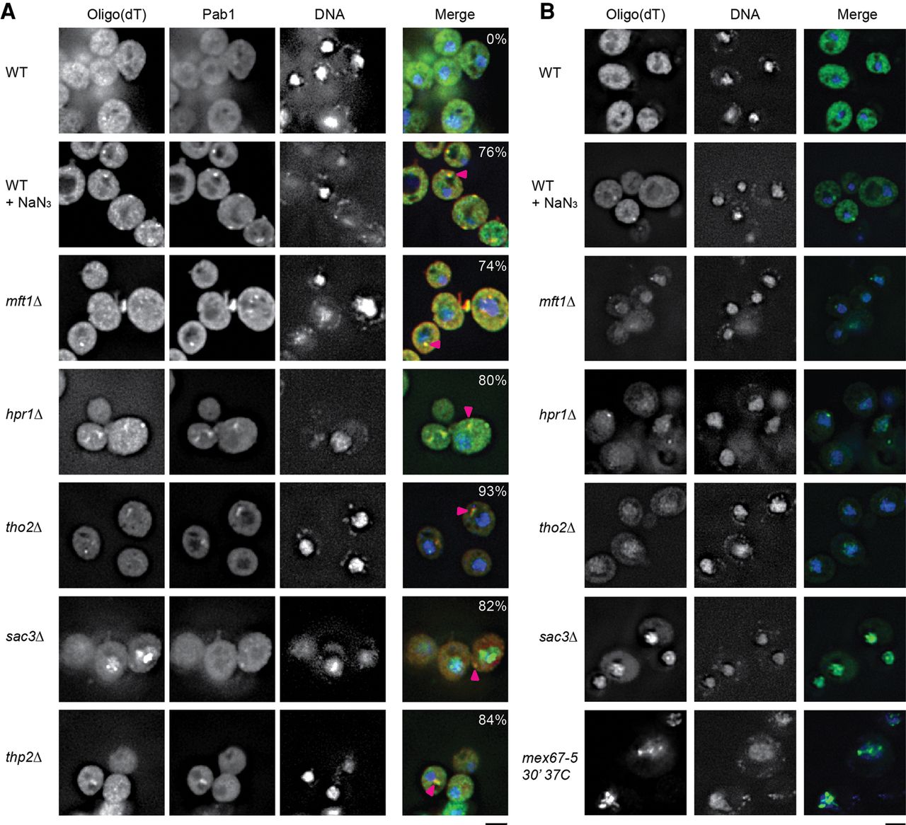
THO and TREX-2 TT foci harbor mRNA. (A) FISH using an oligo(dT) probe, combined with Pab1 immunofluorescence was conducted as detailed in Materials and Methods. Cells were additionally mounted in DAPI-containing media to visualize DNA. WT cells were stressed with NaN3 as previously described. Pink arrowheads indicate colocalization of RNA and Pab1 signal. Percentage values indicate percentage of Pab1 foci overlapping with an oligo(dT) foci. Data representative of observations in two biological replicates (separate culture and transformant). Scale bar = 2.5 µM. (B) FISH was conducted as in A, but without Pab1 immunofluorescence. Notably, the nuclear accumulation of oligo(dT) signal in the nucleus was more apparent, suggesting loss of signal due to the immunofluorescence protocol. Mex67-5 was shifted to the nonpermissive temperature for 30 min prior to fixation. Scale bar = 2.5 µM




