


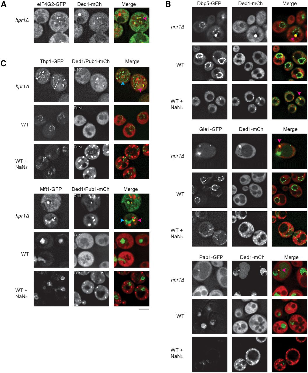
Factors involved in export and 3′ end formation partially colocalize with hpr1Δ TT foci, or exhibit mislocalization in cells with such foci. (A) hpr1Δ eIF4G2-GFP strain transformed with pRP2132 (Ded1-mCh), examined under mid-log conditions. (B) hpr1Δ or WT GFP-tagged strains (Dbp5, Gle1, Pap1) were transformed with pRP2132 (Ded1-mCh) and examined under mid-log conditions ± NaN3 (30 min of 0.5% w/v). (C) As in B except Pub1-mCh (pRP2150) was utilized in place of Ded1-mCh in specific Mft1 or Thp1-GFP-tagged strains as indicated. In all hpr1Δ data sets above, pink arrowheads indicate colocalization of GFP-tagged proteins with Ded1-mCh; blue arrowheads indicate Ded1-distinct GFP foci. Data representative of observations in two biological replicates (separate culture and transformant). Scale bar = 2.5 µM. Colocalization frequencies summarized in Supplemental Table S2.




