


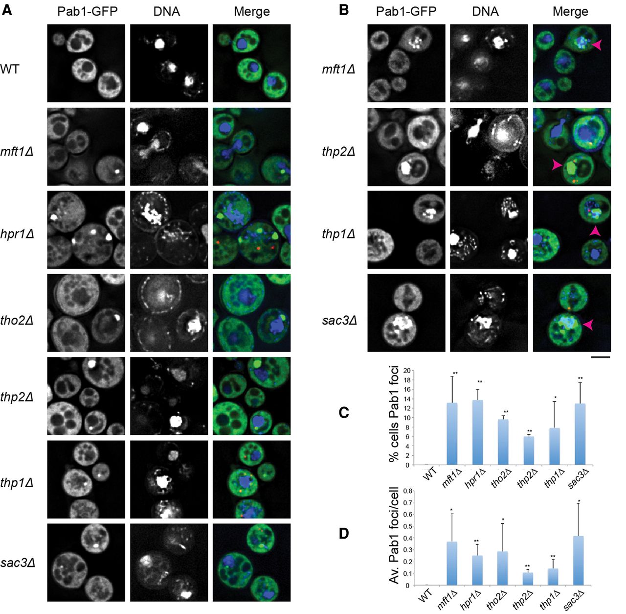
Deletion of THO and TREX-2 complex members leads to constitutive Pab1 foci under nonstress conditions. (A) Strains were transformed with pRP1657 expressing Pab1-GFP (stress granule marker) and Edc3-mCh (P-body marker; shown only in merges due to space constraints), grown to mid-log, incubated with hoescht DNA stain (10 µg/mL for 30 min), and examined. No difference in foci behavior was observed ± hoescht. (B) Examples of nuclear accumulation of Pab1, highlighted by pink arrowheads. Scale bar = 2.5 µM. (C) Average number of cells with Pab1-GFP (“TT”) foci. Data from three biological replicates (separate culture and transformant) with mean ± SD shown. Paired one-tailed Student's t-tests were conducted to assess significance of mutant data relative to WT; (*) P < 0.05, (**) P < 0.01. (D) Average number of Pab1-GFP (“TT”) foci/cell. Data from three biological replicates (separate cultures and transformant) with mean ± SD shown. Paired one-tailed Student's t-tests were conducted to assess significance of mutant data relative to WT; (*) P < 0.05, (**) P < 0.01




