


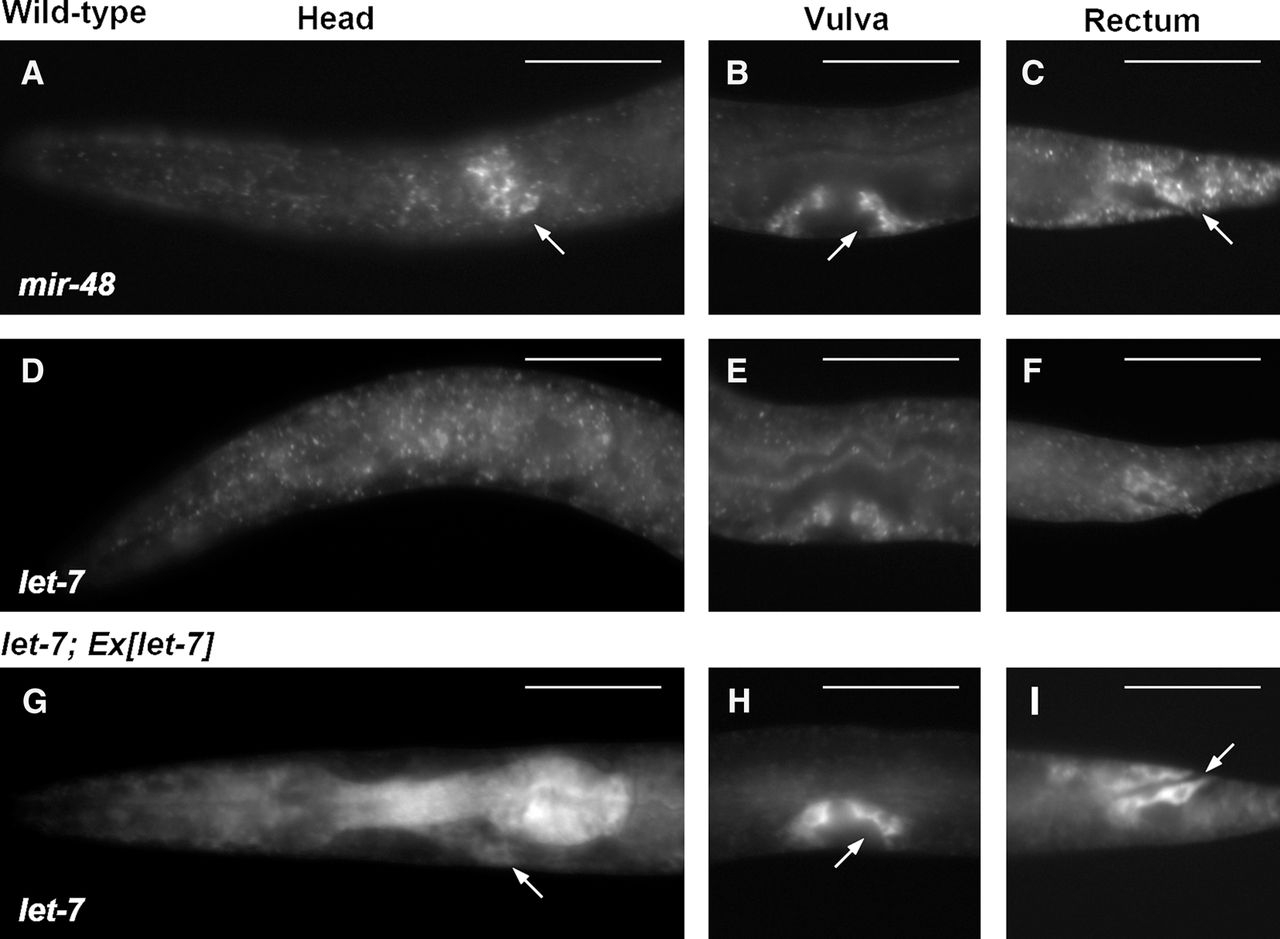
FIGURE 4.
Fluorescent WISH analysis for detection of let-7 family miRNAs at the L4 stage. The family members are mir-48 (A–C) and let-7 (D–I). Genotypes are wild type (A–F) and let-7; Ex[let-7] (G–I). Images show the head region (A,D,G), the vulva (B,E,H), and the rectum (C,F,I). The white arrows in (A–C,G–I) indicate fluorescent signals in the cytoplasm that surround nuclei, which are observed as circles without staining. Color images corresponding to this figure are shown in Supplemental Figure S5. Scale bars, 50 µm.




