


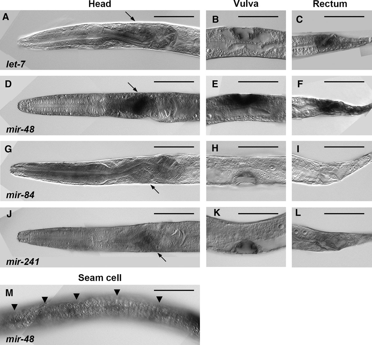
FIGURE 2.
WISH analysis for detection of let-7 family miRNAs at the L4 stage. The family members are let-7 (A–C), mir-48 (D–F,M), mir-84 (G–I), and mir-241 (J–L). Images show the head region (A,D,G,J), the vulva (B,E,H,K), the rectum (C,F,I,L), and seam cells (M). The arrows indicate neurons of the head ganglia (A,D,G,J). The arrowheads point to seam cells (M). Whole-body images corresponding to these partial images are shown in Supplemental Figure S4. Scale bars, 50 µm.




